Unveiling the tumor: a rare case of choroidal melanoma concealed by emulsified silicone oil and retinal detachment
Rami Madani 1,2Jad Madani 3
Adam Ayoub 1
Ali J. Haidar 4
Ben J. Glasgow 5
Daniel B. Rootman 6
Mehran Taban 7
Tarek Alasil 8
1 WMU Homer Stryker M.D. School of Medicine, Department of Surgical Sciences, Kalamazoo, Michigan, United States
2 University of California San Francisco Fresno, Department of Surgery, Fresno, California, United States
3 University of California, Los Angeles, Department of Ecology and Evolutionary Biology, Los Angeles, California, United States
4 University of California, Los Angeles, David Geffen School of Medicine, Los Angeles, California, United States
5 Jules Stein Eye Institute, Department of Pathology, Los Angeles, California, United States
6 Jules Stein Eye Institute, Division of Orbital and Ophthalmic Plastic Surgery, Los Angeles, California, United States
7 Macula Retina Vitreous Center, Los Angeles, California, United States
8 Vitreoretinal Surgery, Retina Innovation Institute, Manhattan Beach, California, United States
Abstract
Objective: To highlight the necessity of maintaining a high index of suspicion for the timely recognition and intervention in patients with choroidal melanoma and exudative retinal detachment masked by emulsified silicone oil.
Methods: Case report
Results: We report a challenging case of a 61-year-old Hispanic male with a history of vitrectomy and silicone oil tamponade for presumed retinal detachment. The examination was notable for elevated intraocular pressure, anterior chamber emulsified silicone oil, and 360-degree posterior synechiae obscuring posterior segment visualization. Initial management targeted silicone oil-induced ocular hypertension, but persistent symptoms prompted surgical intervention. Intraoperatively, a large pigmented choroidal mass with associated exudative retinal detachment was discovered. Subsequent B-scan ultrasonography confirmed a dome-shaped choroidal tumor. Enucleation was performed, and histopathology confirmed choroidal melanoma with coexisting chronic pigmented perisilicone vitreoretinopathy.
Conclusion: This case highlights the importance of maintaining a high index of suspicion for intraocular tumors in patients with unexplained retinal detachments, especially when visualization is hindered by prior vitreoretinal surgery and emulsified silicone oil. Multimodal imaging, including ultrasonography or magnetic resonance imaging, should be considered in cases where clinical findings are incongruent or obscured. Timely recognition and intervention remain critical for optimal oncologic and visual outcomes.
Introduction
Choroidal melanoma is the most common primary intraocular malignancy in adults, arising from melanocytes within the uveal tract. This cancer accounts for approximately 90% of uveal melanomas and shows a strong predilection for individuals of White race, who constitute about 98% of cases. The annual incidence in the United States is estimated at 5–6 cases per million, and the prevalence of precursor choroidal nevus in White adults is about 5% [1], [2].
Key risk factors include White race, light iris color, and increased choroidal pigmentation, with the latter paradoxically associated with higher risk in White populations [3]. There is also a higher prevalence in individuals with multiple dysplastic nevi and a personal or family history of cutaneous melanoma, suggesting shared genetic susceptibility. Female reproductive factors, such as early age at first and last childbirth and higher body mass index in postmenopausal women, are associated with increased risk of choroidal nevus, a precursor lesion, possibly mediated by increased lifetime unopposed estrogen exposure [4].
Early diagnosis and intervention are critical, as delayed treatment worsens prognosis. Diagnosis often relies on multimodal imaging, including ultrasonography, optical coherence tomography (OCT), and fundus autofluorescence [5], [6]. Management strategies for localized disease include plaque brachytherapy, proton beam radiotherapy, transpupillary thermotherapy, local resection, and enucleation [5]. However, advanced or metastatic disease, often refractory to conventional therapies, necessitates systemic treatment approaches that remain largely palliative in nature [2], [7]. Importantly, in vitrectomized eyes containing silicone oil, postoperative findings such as elevated intraocular pressure, anterior chamber oil, or 360-degree posterior synechiae may mimic expected surgical sequelae, leading clinicians to anchor on benign postoperative explanations and delay essential imaging.
This case report presents an unusual instance of small suspicious choroidal lesion initially noted during retinal detachment repair. Poor compliance with follow up after surgery and the pupillary block secondary to silicone oil masked the underlying progression of the tumor, leading to a delay in definitive management.
Case description
A 61-year-old Hispanic man with a history of pars plana vitrectomy and silicone oil tamponade for presumed rhegmatogenous retinal detachment in the left eye, performed one year earlier at an outside institution, was referred to our retina service because of progressive vision loss (hand motion) and ocular pain. At the time of the initial surgery, a small choroidal lesion was noted, raising concern for early choroidal melanoma. Although the patient was referred to ocular oncology, he did not follow up, and the lesion remained unchecked until presentation to our service one year later. On examination, intraocular pressure was elevated to 32 mmHg. Slit-lamp biomicroscopy revealed a reverse hypopyon and 360-degree posterior synechiae, consistent with pupillary block from emulsified silicone oil in the anterior chamber. There was no view to the posterior segment.
The patient was started on maximally tolerated topical therapy, including fixed combination dorzolamide 2%, timolol 0.5% ophthalmic solution twice daily, brimonidine 0.2% twice daily, and prednisolone acetate 1% once daily to address the associated inflammation. Despite adherence to this regimen, intraocular pressure remained elevated, and the patient continued to experience ocular discomfort.
Considering the inadequate response to medical therapy and the pupillary block secondary to anteriorly migrated silicone oil, a decision was made to proceed with surgical intervention. Intraoperatively, posterior synechiolysis and removal of emulsified silicone oil were performed. A large pigmented intraocular mass was identified, arising from the inferior choroid, extending to the macula, and markedly impairing intraoperative visualization with endoillumination. An associated inferior exudative retinal detachment was also observed (Figure 1 [Fig. 1]).
Figure 1: Post-operative B-scan confirmed a large choroidal mass with medium internal reflectivity occupying half of the globe with adjacent exudative retinal detachment (a). The growing choroidal mass pushed the emulsified silicone oil into the anterior chamber and hence contributed to the reverse hypopyon and 360 posterior synechiae. Intraoperative views through the BIOM demonstrated a large choroidal melanoma originating from the inferior choroid, covering the macula, and interfering with the endo-illumination through the pars plana (b, c).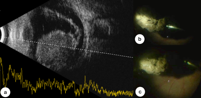
Postoperative B-scan ultrasonography revealed a dome-shaped choroidal mass occupying approximately 50% of the globe, with medium internal reflectivity and an adjacent exudative retinal detachment (Figure 1a [Fig. 1]). Systemic metastatic workup, including computed tomography of the chest abdomen and pelvis and magnetic resonance imaging (MRI) of the brain and orbit, were negative for extraocular metastasis. Given the poor visual prognosis and intraocular tumor diagnosis, a multidisciplinary decision was made to enucleate the affected globe.
Gross examination of the enucleated eye disclosed a pigmented choroidal mass measuring 12 mm in diameter and 11 mm in thickness, along with an associated exudative retinal detachment. Histopathologic analysis confirmed a diagnosis of choroidal melanoma, composed of densely pigmented spindle cells arranged in fascicles with prominent nucleoli (Figure 2 [Fig. 2]). Additionally, extensive fibrovascular membranes with pigment-laden macrophages and vacuolated spaces consistent with chronic pigmented perisilicone vitreoretinopathy were identified, underscoring the prolonged presence and emulsification of intraocular silicone oil (Figure 2 [Fig. 2]).
Figure 2: Stereomicroscopy of the enucleated left eye shows a large, pigmented mass with an extensive exudative retinal detachment (a). Microscopically, densely pigmented spindle cells are arranged in fascicles with prominent nucleoli, indicative of choroidal melanoma (b). In addition, dense fibrovascular membranes bear clusters of pigment-laden macrophages with large vacuoles suggestive of pigmented perisilicone vitreoretinopathy (c).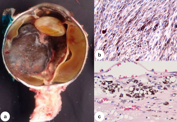
Discussion
This case illustrates a diagnostically challenging presentation of a large choroidal melanoma in a vitrectomized eye with silicone oil tamponade and concurrent exudative retinal detachment (ERD). To our knowledge, there are no published studies reporting a case of choroidal melanoma and ERD concealed by emulsified silicone oil. Patients with choroidal melanoma are often asymptomatic, with an estimated 30% showing no visual complaints at the time of diagnosis. When symptoms are present, they may include blurred vision, photopsia, and floaters, with ocular pain being less frequent secondary to the intraocular tumor itself or its sequelae.
Choroidal melanoma is a well-established cause of ERD, driven by tumor-induced disruption of the retinal pigment epithelium (RPE) and choriocapillaris. The detachment results from increased choroidal vascular permeability and breakdown of the outer blood-retinal barrier, often associated with microvascular loops, networks, and the secretion of angiogenic and permeability factors by tumor and inflammatory cells. Larger tumors with complex vasculature are more likely to exhibit exudation due to extensive RPE compromise. When an underlying tumor is not adequately visualized on examination, the secondary exudative retinal detachment can easily be mistaken for a primary retinal detachment. Recognition of this pathophysiology is critical, particularly in eyes with opaque media or prior surgical intervention, and therefore any unexplained retinal detachment should prompt careful evaluation for an intraocular malignancy [7], [8].
Intraocular silicone oil was utilized in our patient as a tamponade following retinal detachment repair, but this further impeded visualization of the posterior segment. The high refractive index of silicone oil can potentially interfere with ultrasonography [9], [10]. Further, silicone oil has been associated with retinal changes, including preretinal or epiretinal deposits which may mask or mimic various intraocular pathology [11].
Importantly, even in rare cases where B-scan ultrasonography is employed to evaluate obscured posterior structures, tumor detection may be compromised by both the anatomical effects of the ERD and silicone oil interference, underscoring the diagnostic challenges in such complex presentations.
This case underscores the importance of maintaining a high index of suspicion for intraocular malignancy in patients with atypical or unexplained retinal detachments, especially in the context of opaque media or prior vitreoretinal surgery. Preoperative posterior segment visualization was precluded by posterior synechiae and emulsified silicone oil, emphasizing the necessity of B-scan ultrasonography and MRI in such cases to rule out occult intraocular tumors. Routine pre-surgical imaging is critical when clinical findings are incongruent with a primary rhegmatogenous detachment, or when detachment characteristics suggest an exudative etiology. In eyes undergoing silicone oil tamponade, serial imaging may be warranted to detect interval changes indicative of tumor growth. Prompt identification is essential to avoid delayed diagnosis and to guide timely referral for oncologic management.
Notes
Ethics statement
Written informed consent was obtained from the patient for publication of this case report and any accompanying images. Committee ethics approval was not required.
Author contributions
RM and TA contributed to the conception and design of this project. RM performed data extraction and analysis. RM, AA, AJH, and JM conducted the literature review and wrote sections of the manuscript. RM, BJG, DBR, MT, and TA supervised and structured drafting of the manuscript. All authors contributed to the article and approved the submitted version.
Competing interests
The authors declare that they have no competing interests.
References
[1] Shields CL, Manalac J, Das C, Ferguson K, Shields JA. Choroidal melanoma: clinical features, classification, and top 10 pseudomelanomas. Curr Opin Ophthalmol. 2014 May;25(3):177-85. DOI: 10.1097/ICU.0000000000000041[2] Chien JL, Sioufi K, Surakiatchanukul T, Shields JA, Shields CL. Choroidal nevus: a review of prevalence, features, genetics, risks, and outcomes. Curr Opin Ophthalmol. 2017 May;28(3):228-37. DOI: 10.1097/ICU.0000000000000361
[3] Harbour JW, Brantley MA, Hollingsworth H, Gordon M. Association between choroidal pigmentation and posterior uveal melanoma in a white population. Br J Ophthalmol. 2004 Jan;88(1):39-43. DOI: 10.1136/bjo.88.1.39
[4] Qiu M, Shields CL. Relationship between female reproductive factors and choroidal nevus in US women: analysis of data from the 2005-2008 National Health and Nutrition Examination Survey. JAMA Ophthalmol. 2015 Nov;133(11):1287-94. DOI: 10.1001/jamaophthalmol.2015.3178
[5] Shields CL, Lim LS, Dalvin LA, Shields JA. Small choroidal melanoma: detection with multimodal imaging and management with plaque radiotherapy or AU-011 nanoparticle therapy. Curr Opin Ophthalmol. 2019 May;30(3):206-14. DOI: 10.1097/ICU.0000000000000560
[6] Obuchowska I, Konopińska J. Importance of optical coherence tomography and optical coherence tomography angiography in the imaging and differentiation of choroidal melanoma: a review. Cancers (Basel). 2022 Jul 10;14(14):3354. DOI: 10.3390/cancers14143354
[7] Tomkins-Netzer O, Niederer R, Greenwood J, Fabian ID, Serlin Y, Friedman A, Lightman S. Mechanisms of blood-retinal barrier disruption related to intraocular inflammation and malignancy. Prog Retin Eye Res. 2024 Mar;99:101245. DOI: 10.1016/j.preteyeres.2024.101245
[8] Kivelä T, Eskelin S, Mäkitie T, Summanen P. Exudative retinal detachment from malignant uveal melanoma: predictors and prognostic significance. Invest Ophthalmol Vis Sci. 2001 Aug;42(9):2085-93.
[9] Chen Y, Kearns VR, Zhou L, Sandinha T, Lam WC, Steel DH, Chan YK. Silicone oil in vitreoretinal surgery: indications, complications, new developments and alternative long-term tamponade agents. Acta Ophthalmol. 2021 May;99(3):240-50. DOI: 10.1111/aos.14604
[10] Barca F, Caporossi T, Rizzo S. Silicone oil: different physical proprieties and clinical applications. Biomed Res Int. 2014;2014:502143. DOI: 10.1155/2014/502143
[11] Ferrara M, Coco G, Sorrentino T, Jasani KM, Moussa G, Morescalchi F, Dhawahir-Scala F, Semeraro F, Steel DHW, Romano V, Romano MR. Retinal and corneal changes associated with intraocular silicone oil tamponade. J Clin Med. 2022 Sep 5;11(17):5234. DOI: 10.3390/jcm11175234