
Synthese von Silber- und Kupfer-Quantenpunkten mit Eichelextrakt reduziert die Biofilm-Genexpression in klinischen Isolaten von Staphylococcus aureus und Acinetobacter baumannii
Yasman Sadat Nabipour 1Ardeshir Hesampour 1
Salman Ahmady Asbchin 2
Maryam Tajabadi 1
Arman Rostamzad 3
1 Department of Biology, CT.C., Islamic Azad University, Tehran, Iran
2 Department of Microbiology, Faculty of science, University of Mazandaran, Babolsar, Iran
3 Department of Biology, Faculty of science, Ilam University, Ilam, Iran
Abstract
Background: The emergence of multidrug-resistant (MDR) bacteria, particularly Staphylococcus (S.) aureus and Acinetobacter (A.) baumannii, poses a critical global health challenge due to their strong ability to form biofilms. Alternative strategies targeting biofilm formation are urgently needed.
Methods: Silver (Ag) and copper (Cu) quantum dots (QDs) were synthesized via a green hydrothermal method using oak fruit extract. Their antimicrobial and antibiofilm activities were tested against strong biofilm-producing clinical isolates of S. aureus (n=5) and A. baumannii (n=5) using broth microdilution and biofilm inhibition assays. The effect of sub-MIC concentrations of QDs on biofilm-associated genes (icaA, icaD, icaR in S. aureus; bap, ompA in A. baumannii) was evaluated by RT-qPCR.
Results: The MIC values of Ag-QDs and Cu-QDs were 300 µg/mL and 400 µg/mL, respectively. Both QDs significantly inhibited biofilm formation at sub-MIC levels. RT-qPCR analysis showed marked downregulation of all tested genes following QD treatment, with Ag-QDs exhibiting a stronger inhibitory effect than Cu-QDs.
Conclusion: Green-synthesized Ag-QDs and Cu-QDs demonstrate significant antibiofilm activity by suppressing key biofilm-related genes in S. aureus and A. baumannii. These findings highlight their potential as eco-friendly nano-antibiofilm agents for infection control.
Keywords
biofilm inhibition, Staphylococcus aureus, Acinetobacter baumannii, quantum dots, green synthesis
Introduction
The escalating crisis of bacterial antibiotic resistance represents a significant threat to global public health, necessitating the urgent development of novel therapeutic strategies [1], [2]. Among the mechanisms contributing to this resistance, biofilm formation is particularly important, as it confers enhanced protection against antimicrobial agents and host immune responses, rendering infections difficult to eradicate and increasing the risk of healthcare-associated infections (HAIs) [3]. Quantum dots (QDs), as innovative nanomaterials, have emerged as promising tools in this context due to their intrinsic antimicrobial properties, structural stability, and distinct modes of action that may reduce the likelihood of resistance development [4], [5].
QDs are multifunctional materials that have shown remarkable potential in biomedical applications, especially in targeting pathogenic biofilms [6]. Their antimicrobial activity involves multiple mechanisms, including physical disruption of cell membranes, generation of reactive oxygen species (ROS), and, in certain cases, photothermal or photodynamic effects [7]. These mechanisms allow QDs to inhibit a wide spectrum of bacterial species at lower concentrations than many conventional antibiotics, highlighting their potential as alternative or adjunctive therapeutics in infection control [8], [9].
Staphylococcus (S.) aureus, classified by the World Health Organization as a high-priority pathogen, is a major cause of HAIs, particularly due to its ability to form resilient biofilms on medical devices and host tissues [10]. The formation and maturation of S. aureus biofilms are primarily mediated by the polysaccharide intercellular adhesion (PIA) system, encoded by the icaADBC operon [11]. Among the key genes, icaA encodes an N-acetylglucosaminyltransferase essential for polysaccharide synthesis, while icaD acts as a chaperone supporting biofilm formation. Regulatory elements, such as icaR, modulate the expression of these structural genes, influencing the robustness of biofilm development [12], [13].
Acinetobacter (A.) baumannii is a formidable opportunistic pathogen responsible for severe HAIs and is often multidrug-resistant (MDR) [14]. Its pathogenicity is strongly associated with the formation of antimicrobial-resistant biofilms, which facilitate persistent colonization and horizontal transfer of resistance genes [15]. Critical biofilm-associated virulence factors in A. baumannii include the biofilm-associated protein (Bap), which mediates cell-cell adhesion, and the outer membrane protein A (OmpA), which promotes adherence to biotic and abiotic surfaces, thereby contributing to both biofilm integrity and infection persistence [16], [17], [18].
Despite the recognized antimicrobial activity of certain nanoparticles, the specific effects of green-synthesized QDs on biofilm-associated gene regulation in clinical isolates remain largely unexplored [19]. In this study, we synthesized silver and copper QDs using an eco-friendly hydrothermal method with oak fruit extract and evaluated their antibiofilm efficacy against strong biofilm-producing clinical isolates of S. aureus and A. baumannii. Importantly, we aimed to elucidate the molecular mechanisms underlying this activity by assessing the impact of sub-inhibitory concentrations of these QDs on the expression of pivotal biofilm-related genes, including icaA, icaD, and icaR in S. aureus, and bap and ompA in A. baumannii [20], [21]. This approach not only investigates the therapeutic potential of QDs but also provides insights into their role in controlling biofilm-associated virulence, with potential applications in reducing the burden of HAIs and improving infection prevention strategies (Figure 1 [Fig. 1]) [22].
Figure 1: Schematic representation of the green synthesis process of silver (Ag-QDs) and copper (Cu-QDs) quantum dots using oak fruit extract. The extract acts as both reducing and stabilizing agent, leading to nanoparticle formation confirmed by color change and subsequent characterization.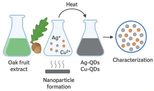
Materials and methods
Bacterial strains and growth conditions
A total of 46 S. aureus and 34 A. baumannii clinical isolates were obtained from diabetic, burn, and surgical wound infections at Razi Hospital between June and December 2023. All isolates were identified using standard biochemical and microbiological tests, including catalase, oxidase, and citrate utilization assays. Reference strains A. baumannii (PTCC 1797) and S. aureus (ATCC 29213) were included as controls. Bacterial cultures were maintained on nutrient agar and stored at 4°C until further use.
Synthesis of quantum dots
Carbon quantum dots (CQDs) were synthesized from oak fruit using a hydrothermal method. Fresh oak fruits were thoroughly washed with deionized water and acetone, air-dried, and ground into a fine powder. Five grams of powder were added to 100 mL deionized water and boiled with vigorous stirring for 30 minutes. The mixture was centrifuged at 5,000 rpm for 15 minutes to obtain a biphasic solution. The supernatant was filtered through Whatman No. 1 filter paper. Formation of CQDs was confirmed via green fluorescence under a TLC-UV chamber. The remaining solution was transferred to an autoclave and heated at 190°C for 5 hours. After cooling to room temperature, the yellow suspension was oven-dried at 80°C for 2 hours to yield a crystalline brown powder, stored at 4°C for further use.
Silver quantum dots (Ag-QDs) were synthesized by adding 20 mL of 1 mM AgNO3 solution dropwise (approximately one drop per second) to 20 mL of oak fruit colloidal solution (0.01 mg/mL) under constant stirring. The reducing agents in the colloid reduced Ag+ to Ag-QDs. After complete addition, the solution was kept in darkness to prevent photochemical reactions. A color change from reddish-brown to yellowish-brown within 1 hour indicated nanoparticle formation. Absorption spectra were recorded at 30°C using a UV-Vis spectrometer. The solution was purified by centrifugation at 10,000 rpm for 10 minutes, and the pellet was dried in an oven at 60°C for 24 hours.
Copper quantum dots (Cu-QDs) were prepared by adding 20 mL of 0.1 M CuSO4 solution to 20 mL of oak fruit extract under continuous stirring at 70°C for 20 minutes. Sodium hydroxide (1 M) was added dropwise to adjust the pH to 12. The solution color transitioned from blue to green, dark green, and finally to dark brown, indicating formation of Cu-based QDs. Brown particles were collected by double centrifugation at 12,000 rpm for 10 minutes each and dried in an oven at 60°C for 24 hours.
Characterization of quantum dots
The absorption spectra of synthesized QDs were recorded using an Agilent UV-Vis spectrometer (200–700 nm) 120 minutes after synthesis (Figure 2 [Fig. 2]).
Figure 2: UV–Vis absorption spectra of silver (Ag-QDs) and copper (Cu-QDs), showing distinct characteristic peaks around 400 nm, confirming the successful synthesis of the quantum dots.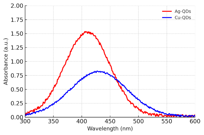
Transmission electron microscopy (TEM): TEM (Zeiss EM10C) was used to determine the morphology and size distribution of the QDs (Figure 3 [Fig. 3]).
Figure 3: TEM micrograph showing spherical morphology and nanoscale distribution of synthesized quantum dots (representative image).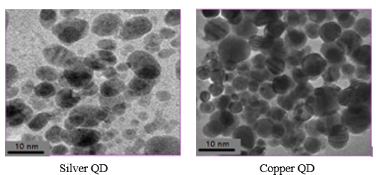
Determination of minimum inhibitory concentration (MIC) and minimum bactericidal concentration (MBC)
MIC and MBC were determined using the broth microdilution method following CLSI guidelines [23]. Briefly, bacterial strains were cultured in Mueller-Hinton broth (MHB) at 37°C for 24 hours with shaking at 120 rpm. Serial dilutions of Ag-QDs and Cu-QDs (50–400 µg/mL) were dispensed into 96-well microplates (50 µL per well), and each well was inoculated with 5 µL of bacterial suspension adjusted to 0.5 McFarland standard. Wells containing bacteria without QDs served as growth controls. MIC was defined as the lowest concentration completely inhibiting visible growth. Sub-MIC concentrations (immediately below MIC) were used for subsequent biofilm and gene expression experiments.
Biofilm formation and antibiofilm assay
Biofilm formation was assessed using the microtiter plate method as described by Lee et al. [21] with optical density (OD) measurements. Each isolate was tested in triplicate. Isolates were classified based on OD values:
- No biofilm producer: OD≤ODc
- Weak biofilm producer: ODc<OD≤2×ODc
- Moderate biofilm producer: 2×ODc<OD≤4×ODc
- Strong biofilm producer: OD>4×ODc.
Five strong biofilm-producing isolates each of S. aureus and A. baumannii were selected for treatment with QDs.
RNA extraction and cDNA synthesis
Total RNA was extracted from isolates treated with sub-MIC concentrations of QDs using a commercial RNA extraction kit (Qiagen, Germany) according to the manufacturer’s protocol. Single colonies were inoculated into 5 mL LB broth and incubated at 37°C for 24 hours. Then, 100 µL of culture were transferred to 10 mL fresh LB broth and grown to mid-lg phase (OD600 ≈0.5) at 37°C with shaking at 185 rpm. RNA quantity and purity were assessed spectrophotometrically at 260, 280, and 230 nm to ensure absence of protein, salt, or other contaminants. Purity ratios (A260/A280 and A260/A230) were within acceptable ranges. Approximately 500 ng of RNA was reverse-transcribed into cDNA using the RT™ Script Prime kit (Takara, Japan).
Quantitative real-ime PCR (qRT-PCR)
qRT-PCR was performed using SYBR Green master mix (Applied Biosystems, UK). Gene-specific primers for bap, ompA ompA, icaA, icaD, and icaR were designed using Primer Express and Gene Runner software, and specificity was verified via NCBI BLAST. The 16S rRNA gene was used as an internal reference. Reactions were prepared in 25 µL volumes, containing 2 µL cDNA template, 10 pM of each forward and reverse primer, 1 µL Bioneer master mix, and nuclease-free water to 25 µL. Amplification was performed on a Bioneer Exicycler 96 with the following thermal profile:
- Initial denaturation: 95°C for 10 min
- 40 cycles: 95°C for 20 s, 58°C for 40 s
- Melting curve: 95°C for 15 s, 60°C for 30 s, 95°C for 15 s
All reactions were performed in duplicate. Relative gene expression was calculated using the 2^–∆∆Ct method. Table 1 [Tab. 1] shows the sequence of forward and reverse primer pairs of bap, ompA, and ica A, D, R genes.
Table 1: Sequences of oligonucleotide primers used for quantitative real-time PCR (qRT-PCR) analysis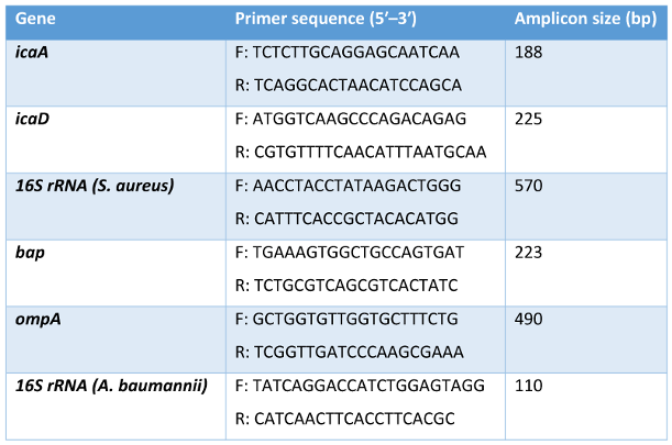
Statistical analysis
All statistical analyses were performed using SPSS version 18 (SPSS Inc., Chicago, IL, USA). Gene expression data were analyzed using one-way ANOVA followed by Tukey’s post hoc test for multiple comparisons. Differences with p-values <0.05 were considered statistically significant.
Results
Biofilm production
Among the clinical isolates screened, 36 S. aureus and 34 A. baumannii strains were evaluated for their biofilm-forming capacity using the microtiter plate assay. Five isolates from each species, identified as strong biofilm producers, were selected for subsequent experiments. Isolates exhibiting weak or moderate biofilm formation were excluded from further analyses to focus on the most virulent biofilm-forming strains. The distribution of biofilm production ability among S. aureus isolates from different clinical samples is detailed in Table 2 [Tab. 2]. Similarly, the biofilm formation capacity of A. baumannii isolates is summarized in Table 3 [Tab. 3].
Table 2: Biofilm production ability of S. aureus isolates from different clinical samples

MIC and sub-MIC of silver and copper QDs
The MIC values for the selected strong biofilm-producing isolates were determined to be 300 µg/mL for Ag-QDs and 400 µg/mL for Cu-QDs for both S. aureus and A. baumannii. Based on these results, sub-MIC concentrations were defined as 200 µg/mL for Ag-QDs and 300 µg/mL for Cu-QDs. These concentrations were used for subsequent antibiofilm and gene expression experiments to assess the effects of QDs without completely inhibiting bacterial growth (Figure 3 [Fig. 3]).
Anti-biofilm activities of silver and copper QDs
Both Ag-QDs and Cu-QDs demonstrated significant antibiofilm activity against all isolates. Complete inhibition of biofilm formation was observed at concentrations ≥300 µg/mL for Ag-QDs and ≥400 µg/mL for Cu-QDs, as indicated by optical density measurements below the cut-off value (ODc=0.22). Reference strains exhibited similar susceptibility patterns, confirming the reproducibility and consistency of the results. Sub-MIC concentrations of QDs significantly reduced biofilm biomass, indicating effective antibiofilm activity even at non-lethal concentrations.
Effect of sub-MIC concentrations of QDs on biofilm-related gene expression: Quantitative real-time PCR analysis revealed that treatment with sub-MIC concentrations of both Ag-QDs and Cu-QDs significantly downregulated the expression of key biofilm-associated genes in both bacterial species. In S. aureus, expression levels of icaA, icaD, and icaR were markedly decreased (p<0.05) following treatment with QDs. Ag-QDs consistently demonstrated a more pronounced inhibitory effect on these genes compared to Cu-QDs. In A. baumannii, bap and ompA gene expression was significantly reduced under sub-MIC QD treatment (p<0.05). Ag-QDs again exhibited stronger suppression of gene expression than Cu-QDs.
Real-time PCR assessment of icaA and icaD expression in S. aureus
The analysis of gene expression revealed a statistically significant decrease in icaA, icaD, and icaR levels in S. aureus following treatment with Ag-QDs and Cu-QDs compared to untreated controls (p<0.05). Notably, Ag-QDs exerted a stronger inhibitory effect on gene expression than Cu-QDs across all tested isolates (Figure 4 [Fig. 4]).
Figure 4: Relative expression of biofilm-related genes (icaA, icaD, icaR in Staphylococcus aureus; bap and ompA in Acinetobacter baumannii) following treatment with sub-MIC concentrations of silver quantum dots (Ag-QDs) and copper quantum dots (Cu-QDs). Expression levels were normalized to 16S rRNA and calculated using the 2^–∆∆Ct method. Both Ag-QDs and Cu-QDs significantly downregulated gene expression compared with untreated controls, with Ag-QDs exhibiting a stronger inhibitory effect (p<0.05)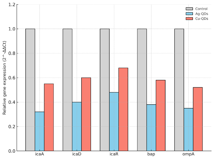
Discussion
A. baumannii represents a particularly formidable pathogen, with its rising prevalence in healthcare settings and extensive drug resistance profiles contributing substantially to patient morbidity and mortality [24], [25], [26]. Despite its recognized clinical significance, the ecology and transmission dynamics of A. baumannii in non-hospital environments remain insufficiently understood [24], [25]. Similarly, S. aureus continues to be a predominant cause of HAIs, with its ability to develop resistance to multiple antibiotics and form biofilms on medical devices posing major therapeutic challenges. Biofilm formation constitutes a critical virulence mechanism, substantially contributing to multidrug resistance [26], [27]. Chronic biofilm-associated infections result in prolonged treatment, increased mortality, and considerable economic burdens, particularly in developing countries, emphasizing the urgent need for alternative therapeutic strategies [28].
Nanotechnology has emerged as a promising approach to address biofilm-related challenges. The application of nanoparticles in antimicrobial research has gained significant attention, resulting in rapid advancement in this field [29]. Among various nanomaterials, quantum dots (QDs) exhibit unique physicochemical properties and multifunctional antimicrobial capabilities, making them particularly suitable for combating biofilm-associated infections [30], [31]. While previous studies and comprehensive reviews have addressed nanoparticle-based antibiofilm strategies, our study specifically investigates the antibiofilm mechanisms of green-synthesized metallic QDs at the genetic level, representing, to our knowledge, the first report on the effects of silver and copper QDs on the expression of key biofilm-related genes in clinical isolates of S. aureus and A. baumannii [29], [30].
In this study, we employed an environmentally friendly synthesis method using oak fruit extract, consistent with the principles of green chemistry [31]. Both Ag-QDs and Cu-QDs synthesized through this approach demonstrated significant antibacterial and antibiofilm activities against clinical isolates of S. aureus and A. baumannii [32]. Importantly, at sub-MIC concentrations, these QDs effectively suppressed the expression of critical biofilm-related genes, with Ag-QDs showing superior efficacy compared to Cu-QDs [25], [28]. The observed MIC values, 300 µg/mL for Ag-QDs and 400 µg/mL for Cu-QDs, are consistent with previous reports indicating the potent antibacterial activity of silver nanoparticles against S. aureus [27], [31].
Our results extend existing knowledge by providing novel insights into the genetic mechanisms underlying QD-mediated biofilm inhibition [33]. Quantitative real-time PCR analysis revealed significant downregulation of icaA, icaD, and icaR genes in S. aureus, and bap and ompA genes in A. baumannii following treatment with sub-MIC concentrations of QDs [34], [35]. These findings suggest that QDs interfere with biofilm formation by disrupting key pathways involved in bacterial adhesion, quorum sensing, and extracellular matrix production. Notably, Ag-QDs consistently demonstrated stronger inhibitory effects on gene expression compared to Cu-QDs, highlighting the higher potency of silver-based nanomaterials in antibiofilm applications [36], [23].
Our findings agree with previous studies showing that metallic nanoparticles are capable of modulating biofilm-associated gene expression. Earlier research has demonstrated that such nanoparticles can alter the expression of genes involved in biofilm formation in S. aureus as well as reduce the expression of key biofilm-related genes in A. baumannii [31], [32], [34]. Other studies have also indicated that treatment with metallic nanoparticles leads to a simultaneous decrease in virulence gene expression and biofilm production in multidrug-resistant clinical strains [18]. The present study confirms and extends these observations by showing that both silver and copper QDs, even at sub-MIC concentrations, exert similar inhibitory effects, supporting their potential as eco-friendly antibiofilm agents against clinical isolates [37], [38].
The correlation between reduced biofilm formation and downregulation of key virulence genes in both bacterial species provides mechanistic insight into QD-mediated antibiofilm activity. The suppression of icaADR genes in S. aureus and bap/ompA genes in A. baumannii explains the observed reduction in biofilm biomass and highlights the potential of QDs to penetrate biofilm structures and interfere with critical molecular pathways [26], [30]. These findings not only reinforce the potential of metallic QDs as alternative therapeutic agents against resistant pathogens but also suggest a promising strategy for overcoming biofilm-associated antibiotic resistance in clinical settings [4], [29].
In summary, this study provides evidence that green-synthesized Ag-QDs and Cu-QDs exert significant antibiofilm activity against clinical isolates of S. aureus and A. baumannii through downregulation of key genetic determinants of biofilm formation, even at sub-inhibitory concentrations. Among the two, Ag-QDs demonstrated consistently superior efficacy, supporting their preferential consideration for further development.
Limitations
However, several critical aspects must be addressed before clinical translation. First, while our in vitro findings are mechanistically informative, they do not account for the complex host–pathogen–nanomaterial interactions present in vivo. Second, the long-term biocompatibility and potential cytotoxicity of these QDs, particularly upon repeated administration, remain to be systematically evaluated. Third, the emergence of bacterial tolerance to sub-MIC levels of QDs, though not observed in our short-term assays, warrants investigation through longitudinal exposure studies. From a translational perspective, future research should prioritize in vivo efficacy testing in appropriate animal models, pharmacokinetic and biodistribution profiling, and formulation development to enhance targeted delivery to biofilm-infected tissues. Additionally, combination strategies with conventional antibiotics or biofilm-disrupting enzymes could be explored to achieve synergistic effects and reduce the risk of resistance. Ultimately, addressing these knowledge gaps will be essential to determine whether QD-based therapies can be safely and effectively integrated into clinical practice as adjuncts or alternatives to current antibiotic regimens [7], [15], [20].
Conclusions
This study demonstrates that green-synthesized silver and copper quantum dots exhibit potent antibiofilm activity against clinical isolates of S. aureus and A. baumannii. The observed antibiofilm effects are mediated, at least in part, through the downregulation of key virulence genes involved in biofilm formation, specifically the icaADR operon in S. aureus and bap/ompA genes in A. baumannii. Silver QDs consistently showed superior efficacy compared to copper QDs in suppressing both biofilm formation and virulence gene expression. These findings underscore the potential of metallic quantum dots as promising anti-biofilm agents for combating infections caused by multidrug-resistant pathogens. Future studies should investigate additional genes involved in biofilm regulation and assess the in vivo efficacy and safety of these nanoparticles.
Notes
Competing interests
The authors declare that they have no competing interests.
Authors’ ORCIDs
- Nabipour YS: https://orcid.org/0000-0003-0504-586
- Hesampour A : https://orcid.org/0000-0002-7324-5469
- Asbchin SA : https://orcid.org/0000-0001-6079-1842
- Tajabadi M : https://orcid.org/0000-0003-2780-3447
- Rostamza A : https://orcid.org/0000-0002-2288-3794
Ethical approval
Animal experiments were conducted following the ethical guidelines approved by the Animal Ethics Committee of Tehran University of Medical Sciences (Ethics code: IR.ILAM.REC.1401.008). All in vivo procedures were performed in accordance with the National Institutes of Health (NIH) Guide for the Care and Use of Laboratory Animals and the national guidelines of Iran for the ethical use of animals in research.
Funding
None.
References
[1] Nguyen MH, Le AT, Pham VD, Pham HM, Do HT, Le DT, et al. A Comprehensive study on the antibacterial activities of carbon quantum dots derived from orange juice against Escherichia coli. Appl Sci. 2024 March 16;14(6):2509. DOI:10.3390/app14062509[2] Koul K, Jawanda IK, Soni T, Singh P, Sharma D, Kumari S. Quantum dots: a next generation approach for pathogenic microbial biofilm inhibition; mechanistic insights, existing challenges, and future potential. Arch Microbiol. 2024 Mar;206(4):158. DOI: 10.1007/s00203-024-03919-3
[3] Cepas V, Soto SM. Relationship between biofilm formation and antimicrobial resistance in Gram-negative bacteria. Microb Drug Resist. Feb 9; 2020;26(8):1017-33. DOI:10.1089/mdr.2019.0173
[4] Khatoon Z, McTiernan CD, Suuronen EJ, Mah TF, Alarcon EI. Bacterial biofilm formation on implantable devices and approaches to its treatment and prevention. Heliyon. 2018 Dec;4(12):e01067. DOI: 10.1016/j.heliyon.2018.e01067
[5] Costa MO, Beltrame CO, Ferreira FA, Botelho AM, Lima NC, Souza RC, de Almeida LG, Vasconcelos AT, Nicolás MF, Figueiredo AM. Complete Genome Sequence of a Variant of the Methicillin-Resistant Staphylococcus aureus ST239 Lineage, Strain BMB9393, Displaying Superior Ability To Accumulate ica-Independent Biofilm. Genome Announc. 2013 Aug;1(4):. DOI: 10.1128/genomeA.00576-13
[6] Abd El-Rahman OA, Rasslan F, Hassan SS, Ashour HM, Wasfi R. The RND Efflux Pump Gene Expression in the Biofilm Formation of. Antibiotics (Basel). 2023 Feb;12(2):. DOI: 10.3390/antibiotics12020419
[7] Brossard KA, Campagnari AA. The Acinetobacter baumannii biofilm-associated protein plays a role in adherence to human epithelial cells. Infect Immun. 2012 Jan;80(1):228-33. DOI: 10.1128/IAI.05913-11
[8] McConnell MJ, Actis L, Pachón J. Acinetobacter baumannii: human infections, factors contributing to pathogenesis and animal models. FEMS Microbiol Rev. 2013 Mar;37(2):130-55. DOI: 10.1111/j.1574-6976.2012.00344.x
[9] Hosnedlova B, Kabanov D, Kepinska M, B Narayanan VH, Parikesit AA, Fernandez C, Bjørklund G, Nguyen HV, Farid A, Sochor J, Pholosi A, Baron M, Jakubek M, Kizek R. Effect of Biosynthesized Silver Nanoparticles on Bacterial Biofilm Changes in and. Nanomaterials (Basel). 2022 Jun;12(13):. DOI: 10.3390/nano12132183
[10] Al-Wrafy FA, Al-Gheethi AA, Ponnusamy SK, Noman EA, Fattah SA. Nanoparticles approach to eradicate bacterial biofilm-related infections: A critical review. Chemosphere. 2022 Feb;288(Pt 2):132603. DOI: 10.1016/j.chemosphere.2021.132603
[11] Wu J, Zhang B, Lin N, Gao J. Recent nanotechnology-based strategies for interfering with the life cycle of bacterial biofilms. Biomater Sci. 2023 Feb;11(5):1648-1664. DOI: 10.1039/d2bm01783k
[12] Afrasiabi S, Partoazar A. Targeting bacterial biofilm-related genes with nanoparticle-based strategies. Front Microbiol. 2024;15:1387114. DOI: 10.3389/fmicb.2024.1387114
[13] Ahuatzin-Flores OE, Torres E, Chávez-Bravo E. A Multidrug-Resistant Opportunistic Pathogen in New Habitats: A Systematic Review. Microorganisms. 2024 Mar;12(4):. DOI: 10.3390/microorganisms12040644
[14] Pourkhosravani E, Dehghan Nayeri F, Mohammadi Bazargani M. Decoding antibacterial and antibiofilm properties of cinnamon and cardamom essential oils: a combined molecular docking and experimental study. AMB Express. 2021 Oct;11(1):143. DOI: 10.1186/s13568-021-01305-6
[15] Asadi S, Nayeri-Fasaei B, Zahraei-Salehi T, Yahya-Rayat R, Shams N, Sharifi A. Antibacterial and anti-biofilm properties of carvacrol alone and in combination with cefixime against Escherichia coli. BMC Microbiol. 2023 Mar;23(1):55. DOI: 10.1186/s12866-023-02797-x
[16] Wang Z, Shen Y, Haapasalo M. Antimicrobial and Antibiofilm Properties of Bioceramic Materials in Endodontics. Materials (Basel). 2021 Dec;14(24):. DOI: 10.3390/ma14247594
[17] Kakavan M, Gholami M, Ahanjan M, Ebrahimzadeh MA, Salehian M, Roozbahani F, Goli HR. Expression of bap gene in multidrug-resistant and biofilm-producing Acinetobacter baumannii clinical isolates. BMC Microbiol. 2025 Mar;25(1):108. DOI: 10.1186/s12866-025-03806-x
[18] Hetta HF, Al-Kadmy IMS, Khazaal SS, Abbas S, Suhail A, El-Mokhtar MA, Ellah NHA, Ahmed EA, Abd-Ellatief RB, El-Masry EA, Batiha GE, Elkady AA, Mohamed NA, Algammal AM. Antibiofilm and antivirulence potential of silver nanoparticles against multidrug-resistant Acinetobacter baumannii. Sci Rep. 2021 May;11(1):10751. DOI: 10.1038/s41598-021-90208-4
[19] Rezania N, Rahmati P, Noorbakhsh F, Farhadyar N, Lotfali E. Investigation the effects of silver nanoparticles and gold nanoparticles on expression of and genes in biofilm formation of. Iran J Microbiol. 2022 Aug;14(4):510-517. DOI: 10.18502/ijm.v14i4.10237
[20] Valadbeigi H, Sadeghifard N, Kaviar VH, Haddadi MH, Ghafourian S, Maleki A. Effect of ZnO nanoparticles on biofilm formation and gene expression of the toxin-antitoxin system in clinical isolates of Pseudomonas aeruginosa. Ann Clin Microbiol Antimicrob. 2023 Oct;22(1):89. DOI: 10.1186/s12941-023-00639-2
[21] Lee B, Schjerling CK, Kirkby N, Hoffmann N, Borup R, Molin S, Høiby N, Ciofu O. Mucoid Pseudomonas aeruginosa isolates maintain the biofilm formation capacity and the gene expression profiles during the chronic lung infection of CF patients. APMIS. 2011 Apr;119(4-5):263-74. DOI: 10.1111/j.1600-0463.2011.02726.x
[22] Balouiri M, Sadiki M, Ibnsouda SK. Methods for evaluating antimicrobial activity: A review. J Pharm Anal. 2016 Apr;6(2):71-79. DOI: 10.1016/j.jpha.2015.11.005
[23] Nabipour YS, Hesampour A, Ahmady Asbchin S, Tajabadi M, Rostamzad A. Comparative study of the effects of biologically synthesized copper and zinc oxide quantum nanodots on biofilm-forming genes in Klebsiella pneumoniae and Staphylococcus aureus bacteria. Med Sci J Islamic Azad Univ. 2026; 36 (2) :189-99. DOI: 10.66224/iau.36.2.189
[24] Shawky M, Kalaba MH, El-Sherbiny GM. Tackling carbapenem-resistant (CRAB) and their virulence factors using biosynthesized silver nanoparticles combined with imipenem. Biotechnol Notes. 2025;6:183-195. DOI: 10.1016/j.biotno.2025.07.002
[25] Thilakavathy P, Priyan RM, Jagatheeswari PA, Charles J, Dhanalakshmi V, Lallitha S, Rajendran T, Divya B. Evaluation of Ica Gene in Comparison with Phenotypic Methods for Detection of Biofilm Production by Coagulase Negative Staphylococci in a Tertiary Care Hospital. J Clin Diagn Res. 2015 Aug;9(8):DC16-9. DOI: 10.7860/JCDR/2015/11725.6371
[26] Tyner H, Patel R. Propionibacterium acnes biofilm - A sanctuary for Staphylococcus aureus? Anaerobe. 2016 Aug;40:63-7. DOI: 10.1016/j.anaerobe.2016.05.014
[27] Manandhar S, Singh A, Varma A, Pandey S, Shrivastava N. Biofilm Producing Clinical Isolates Augmented Prevalence of Antibiotic Resistant Cases in Tertiary Care Hospitals of Nepal. Front Microbiol. 2018;9:2749. DOI: 10.3389/fmicb.2018.02749
[28] Rezania N, Rahmati P, Noorbakhsh F, Farhadyar N, Lotfali E. Investigation the effects of silver nanoparticles and gold nanoparticles on expression of and genes in biofilm formation of. Iran J Microbiol. 2022 Aug;14(4):510-517. DOI: 10.18502/ijm.v14i4.10237
[29] Hamida RS, Ali MA, Goda DA, Khalil MI, Al-Zaban MI. Novel Biogenic Silver Nanoparticle-Induced Reactive Oxygen Species Inhibit the Biofilm Formation and Virulence Activities of Methicillin-Resistant (MRSA) Strain. Front Bioeng Biotechnol. 2020;8:433. DOI: 10.3389/fbioe.2020.00433
[30] Heine N, Doll-Nikutta K, Stein F, Jakobi J, Ingendoh-Tsakmakidis A, Rehbock C, Winkel A, Barcikowski S, Stiesch M. Anti-biofilm properties of laser-synthesized, ultrapure silver-gold-alloy nanoparticles against Staphylococcus aureus. Sci Rep. 2024 Feb;14(1):3405. DOI: 10.1038/s41598-024-53782-x
[31] Fareid MA, El-Sherbiny GM, Askar AA, Abdelaziz AM, Hegazy AM, Ab Aziz R, Hamada FA. Impeding Biofilm-Forming Mediated Methicillin-Resistant and Virulence Genes Using a Biosynthesized Silver Nanoparticles-Antibiotic Combination. Biomolecules. 2025 Feb;15(2):. DOI: 10.3390/biom15020266
[32] Mohanta YK, Biswas K, Jena SK, Hashem A, Abd Allah EF, Mohanta TK. Anti-biofilm and Antibacterial Activities of Silver Nanoparticles Synthesized by the Reducing Activity of Phytoconstituents Present in the Indian Medicinal Plants. Front Microbiol. 2020;11:1143. DOI: 10.3389/fmicb.2020.01143
[33] Pal S, Tak YK, Song JM. Does the antibacterial activity of silver nanoparticles depend on the shape of the nanoparticle? A study of the Gram-negative bacterium Escherichia coli. Appl Environ Microbiol. 2007 Mar;73(6):1712-20. DOI: 10.1128/AEM.02218-06
[34] Grygiel I, Bajrak O, Wójcicki M, Krusiec K, Jończyk-Matysiak E, Górski A, Majewska J, Letkiewicz S. Comprehensive Approaches to Combatting Biofilms: From Biofilm Structure to Phage-Based Therapies. Antibiotics (Basel). 2024 Nov;13(11):. DOI: 10.3390/antibiotics13111064
[35] Lin F, Wang Z, Wu FG. Carbon Dots for Killing Microorganisms: An Update since 2019. Pharmaceuticals (Basel). 2022 Oct;15(10):. DOI: 10.3390/ph15101236
[36] He L, Li Z, Gu M, Li Y, Yi C, Jiang M, Yu X, Xu L. Intelligent Carbon Dots with Switchable Photo-Activated Oxidase-Mimicking Activity and pH Responsive Antioxidant Activity Adaptive to the Wound Microenvironment for Selective Antibacterial Therapy. Adv Sci (Weinh). 2024 Oct;11(40):e2406681. DOI: 10.1002/advs.202406681
[37] Nabipour YS, Hesampour A, Asbchin SA, Tajabadi M, Rostamzad A. Green-synthesized quantum dots from quercus brantii for infected polymicrobial wound healing: mechanisms and biocompatibility. BMC Biotechnol. 2026 Jun;:. DOI: 10.1186/s12896-026-01183-5
[38] Swebocki T, Barras A, Kocot A, Płotka M, Boukhterroub R. Minimum inhibitory concentration (MIC) and minimum bactericidal concentration (MBC) assays using broth microdilution method. protocols.io. 2023 Jun 23: 1-12. DOI: 10.17504/protocols.io.5qpvo3x6dv4o/v1