
Green synthesis of carbon quantum dots (CQDs) from oak fruit: antimicrobial and anti-biofilm efficacy against ESKAPE pathogens in wound infections
Yasman Sadat Nabipour 1Ardeshir Hesampour 1
Salman Ahmady Asbchin 2
Maryam Tajabadi 1
Arman Rostamza 3
1 Department of Biology, CT.C., Islamic Azad University, Tehran, Iran
2 Department of Microbiology, Faculty of Science, University of Mazandaran, Babolsar, Iran
3 Department of Biology, Faculty of Science, Ilam University, Ilam, Iran
Abstract
Aim: The present study aims to synthesize Carbon Quantum Dots (CQDs) using natural oak fruit as a green carbon precursor and to evaluate their antimicrobial and anti-biofilm activities against WHO priority antibiotic-resistant ESKAPE pathogens isolated from clinical wound infections.
Methods: CQDs were synthesized via a one-step hydrothermal method (190°C for 5 hours). The study design consisted of four main phases: nanoparticle synthesis and characterization (TEM, FTIR, UV-Vis, fluorescence spectroscopy), in vitro microbiological evaluations (disk diffusion, minimal inhibition concentration/ minimal bactericidal concentration (MIC/MBC), and microtiter plate assays), molecular investigations (Real-Time PCR targeting biofilm-associated genes), and in vivo animal model studies using murine infected wound model. A total of 122 clinical isolates from diabetic, burn, and surgical wounds were collected over 6 months, alongside standard bacterial strains.
Results: Successful synthesis of CQDs was confirmed by blue/green luminescence under 365 nm UV irradiation and the presence of surface functional groups (OH, COOH, C=O) via FTIR. TEM revealed uniform spherical morphology with an average particle size <10 nm. Molecular analysis showed that sub-MIC concentrations of CQDs significantly down-regulated key biofilm-associated genes (icaA/icaD in Staphylococcus aureus, lecA/pelA in Pseudomonas aeruginosa, mrkA/mrkD in Klebsiella pneumoniae, and bap/ompA in Acinetobacter baumannii) compared to the 16S rRNA reference gene (P<0.05). In vivo evaluation demonstrated that topical application of CQDs significantly accelerated epithelialization, improved wound contraction, and reduced wound area over 14 days compared to untreated and antibiotic-treated controls (P<0.05).
Conclusion: Hydrothermally synthesized oak-derived CQDs effectively disrupt biofilm formation at the transcriptional level, reduce wound bacterial burden, and accelerate tissue regeneration. These findings highlight the potential of green-synthesized CQDs as alternative therapeutic agents for managing multidrug-resistant wound infections.
Keywords
antimicrobial resistance, biofilm, carbon quantum dots (CQDs), ESKAPE pathogens, Real-Time PCR, wound healing
Introduction
Antimicrobial resistance (AMR) has emerged as one of the most formidable global public health threats of the 21st century, often described as a “silent pandemic” [1]. The progressive ability of microorganisms to withstand previously effective antimicrobial agents is largely driven by the overuse and misuse of antibiotics in clinical, veterinary, and agricultural settings [2]. In 2019, AMR was directly responsible for over 1.2 million global deaths, and epidemiological models project this toll could escalate to approximately 10 million annual deaths by 2050 if left unaddressed [3]. Among the most critical challenges are the ESKAPE pathogens (Enterococcus faecium, Staphylococcus (S.) aureus, Klebsiella (K.) pneumoniae, Acinetobacter (A.) baumannii, Pseudomonas (P.) aeruginosa, and Enterobacter spp.), which are notorious for their multidrug resistance and are frequently isolated from severe clinical wound infections [4], [5].
A primary mechanism exacerbating bacterial resistance in wound environments is the formation of biofilms. Biofilms are complex, surface-attached microbial communities encased within a self-produced extracellular polymeric substance (EPS) matrix [6]. This protective barrier significantly limits the penetration of conventional antibiotics and shields the embedded bacteria from the host’s immune system [7]. Consequently, biofilm-associated infections, particularly in diabetic, burn, and surgical wounds, lead to prolonged hospitalization, increased healthcare costs, and higher mortality rates [8]. Overcoming the structural and functional barriers of bacterial biofilms remains a critical unmet clinical need [9].
To address these limitations, nanotechnology has been introduced as a highly innovative and promising strategy [10]. By converging phytochemistry and nanophysics, researchers have developed nanoscale materials capable of penetrating the dense biofilm matrix [11]. Quantum Dots (QDs), in particular, have garnered significant attention due to their ultra-small size (typically <10 nm), spherical morphology, and exceptional physicochemical stability [12]. Furthermore, transitioning towards “green synthesis” methods—which utilize natural plant extracts or biomass as reducing and capping agents—offers a sustainable, eco-friendly alternative to traditional chemical syntheses, significantly minimizing reliance on toxic solvents and enhancing biocompatibility for biomedical applications [13], [14].
Carbon quantum dots (CQDs) have recently emerged as a subclass of nanomaterials with low toxicity, high water solubility, and tunable photoluminescence [15]. Several studies have reported the antibacterial activity of CQDs derived from various biomass sources, including orange peel, garlic, and tea leaves [16], [17]. However, the use of oak fruit (Quercus spp.) as a carbon precursor remains largely unexplored. Oak fruit is rich in polyphenols, tannins, and carbohydrates, which can serve as natural reducing and capping agents [18]. Moreover, the anti-biofilm and in vivo wound healing potential of oak-derived CQDs against clinically relevant ESKAPE isolates has not been systematically investigated.
Therefore, the primary aim of this research was to synthesize and characterize oak-derived CQDs and comprehensively evaluate their antibacterial and anti-biofilm efficacy against clinical isolates of ESKAPE pathogens. Additionally, this study sought to investigate the molecular mechanisms underlying biofilm inhibition via Real-Time PCR (targeting specific biofilm-associated genes) and to rigorously assess the in vivo therapeutic potential of these nanoparticles in accelerating tissue regeneration within a murine infected wound model [19].
Materials and methods
Green synthesis and characterization of carbon quantum dots (CQDs)
Carbon quantum dots (CQDs) were synthesized using a one-step, bottom-up hydrothermal method as previously described with modifications [20]. Oak fruit (Quercus robur) was sourced locally from the Mazandaran region, Iran. The fruit was washed thoroughly with deionized water, dried at 80°C for 24 hours, and pulverized into a fine powder using a laboratory grinder. Subsequently, 5 g of the powder was dispersed in 50 mL of deionized water under vigorous stirring for 30 minutes. The mixture was transferred to a 100 mL Teflon-lined stainless-steel autoclave and heated at 190°C for 5 hours. After cooling to room temperature, the resulting dark brown solution was centrifuged at 12,000 rpm for 20 minutes to remove large aggregates and then filtered through a 0.22 µm syringe filter.
The synthesized CQDs were characterized using several analytical techniques. Optical properties were assessed via UV-Vis absorption spectroscopy (Shimadzu UV-1800) and fluorescence spectroscopy (PerkinElmer LS55). The morphology, size, and dispersion of the nanoparticles were determined by transmission electron microscopy (TEM, Zeiss EM10C operating at 80 kV). The presence of surface functional groups was confirmed using Fourier-transform infrared spectroscopy (FTIR, Bruker Tensor 27) in the range of 400–4,000 cm–1 [21].
Bacterial strains and clinical isolates
A total of 122 clinical isolates belonging to the ESKAPE group of pathogens (S. aureus, n=32; K. pneumoniae, n=30; A. baumannii, n=28; P. aeruginosa, n=32) were collected over a six-month period from the central laboratory of Razi Hospital, Tehran, Iran. Samples were obtained from patients with diabetic foot ulcers (n=45), burn wounds (n=40), and surgical site infections (n=37). In addition to the clinical isolates, standard reference strains of the same bacterial species (S. aureus ATCC 25923, K. pneumoniae ATCC 700603, A. baumannii ATCC 19606, P. aeruginosa ATCC 27853) were used as controls. All isolates were cultured on tryptic soy broth (TSB) and Mueller-Hinton Agar (MHA) and incubated at 37°C for 24 hours [22].
In vitro microbiological assays
Initial antibiotic resistance profiles of all isolates were determined using the standard Kirby-Bauer disk diffusion method according to CLSI guidelines (M07-A11,2018) [23]. The capacity of each isolate to form biofilm was quantified using the crystal violet microtiter plate (MTP) assay as previously described [24]. Isolates classified as weak or non-biofilm producers (OD<0.1) were excluded from subsequent experiments.
The minimum inhibitory concentration (MIC) and minimum bactericidal concentration (MBC) of the CQDs were determined using the broth microdilution method in 96-well plates according to CLSI guidelines [23]. Bacterial suspensions (1.5 × 10⁸ colony forming units (CFU)/mL) were exposed to serial dilutions of the nanoparticles (0–500 µg/mL) and incubated for 24 hours at 37°C. The MIC was defined as the lowest concentration with no visible growth, and the MBC was determined by subculturing 10 µL from clear wells onto MHA plates [25].
The anti-biofilm potential of CQDs was assessed by measuring their ability to both inhibit the formation of new biofilms and eradicate pre-formed, mature biofilms (24 h old) using the MTP assay. For inhibition, bacteria were co-incubated with sub-MIC concentrations of CQDs. For eradication, pre-formed biofilms were treated with increasing concentrations of CQDs for 24 hours. Biofilm mass was quantified by crystal violet staining [26].
To investigate the effect of sub-MIC concentrations (1/2 MIC) of CQDs on the expression of key biofilm-associated gene, total RNA was extracted from treated and untreated bacterial cells using TRIzol reagent (Invitrogen) after 12 hours of exposure. RNA quality and quantity were assessed by NanoDrop 2000. cDNA synthesis was performed using a High-Capacity cDNA Reverse Transcription Kit (Applied Biosystems, Foster City, CA, USA). RT-qPCR was carried out using SYBR Green Master Mix (Bio-Rad, Hercules, CA, USA) on a StepOnePlus Real-Time PCR system (Applied Biosystems, Foster City, CA, USA). The following target genes were selected: icaA and icaD in S. aureus; lecA and pelA in P. aeruginosa; mrkA and mrkD in K. pneumoniae; and bap and ompA in A. baumannii. The 16S rRNA gene was used as the endogenous reference control. Relative gene expression was calculated using the 2–ΔΔCt method. All experiments were performed in triplicate [27], [28].
In vivo wound healing study
Animal experiments were conducted following the ethical guidelines approved by the Animal Ethics Committee of Tehran University of Medical Sciences (Ethics code: IR.TUMS.AEC.1402.089). Sixty healthy male BALB/c mice (6–8 weeks old, 20–25 g) were used. A full-thickness excisional wound model (1 cm diameter) was established on the dorsum of each mouse under general anesthesia (ketamine/xylazine). Wounds were subsequently inoculated with 50 µL of a standard concentration (1.5 × 10⁸ CFU/mL) of the respective ESKAPE pathogens. Mice were randomly divided into five groups (n=12 per group): (1) untreated control (no treatment), (2) antibiotic-treated (ciprofloxacin 1% ointment), (3) CQD-treated (1% CQD gel), (4) blank gel control, and (5) sham (no infection, no treatment). Topical treatments were applied daily for 14 days. Wound healing was monitored every 2 days by measuring wound area using digital calipers. On day 14, tissue samples were collected for histopathological analysis (HE staining) and for determining the bacterial load (CFU/g tissue) [29], [30].
Statistical analysis
All experiments were performed in triplicate. Data were expressed as mean ± standard deviation (SD). Statistical analyses were performed using GraphPad Prism 9.0. Comparisons between two groups were made using Student’s t-test, and multiple comparisons were made using one-way ANOVA followed by Tukey’s post-hoc test. A p-value of less than 0.05 was considered statistically significant [31].
Results
Synthesis and characterization of carbon quantum dots (CQDs)
The successful hydrothermal synthesis of CQDs from oak fruit extract was initially indicated by a distinct color change of the reaction mixture from pale yellow to a dark, yellowish-brown hue. Under ultraviolet (UV) irradiation at 365 nm, the synthesized CQDs exhibited strong blue/green fluorescence, confirming the formation of quantum dots with distinct optical properties. The UV-Vis absorption spectrum showed a characteristic peak at 280 nm, corresponding to the π–π* transition of C=C bonds. The fluorescence emission spectrum exhibited excitation-dependent behavior with maximum emission at 450 nm when excited at 360 nm (Figure 1 [Fig. 1]).
Figure 1: UV–Vis absorption spectra of Quercus brantii-mediated quantum dots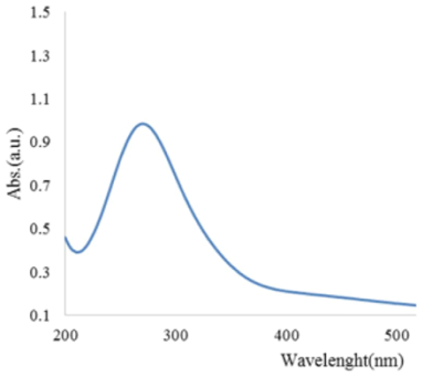
FTIR analysis revealed the presence of surface functional groups: a broad peak at 3,420 cm–1 (O–H stretching), peaks at 2,925 and 2,854 cm–1 (C–H stretching), a sharp peak at 1,635 cm–1 (C=O stretching), and peaks at 1,400 cm–1 and 1,080 cm–1 (C–O and C–O–C stretching). These functional groups indicate high hydrophilicity and excellent dispersibility in aqueous media.
Morphological analysis using transmission electron microscopy (TEM) revealed that the synthesized CQDs possessed a spherical shape with uniform dispersion and no significant agglomeration (Figure 2 [Fig. 2]). The average particle size was determined to be 6.8±1.2 nm (n=100), which is highly favorable for penetrating bacterial biofilms.
Figure 2: (TEM) analysis of Quercus brantii-mediated quantum dots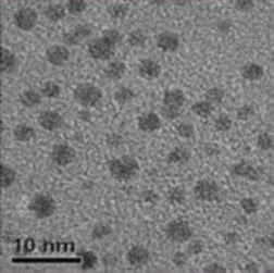
In vitro antimicrobial activity
The antibacterial efficacy of the synthesized CQDs was evaluated against all clinical isolates and standard strains. The MIC values ranged from 125 to 250 µg/mL depending on the bacterial species (Table 1 [Tab. 1], Figure 3 [Fig. 3]). S. aureus exhibited the highest susceptibility (MIC=125 µg/mL for most isolates), whereas P. aeruginosa demonstrated the lowest susceptibility (MIC=250 µg/mL for >70% of isolates). The MBC values were generally 2-fold higher than MIC values. The MBC/MIC ratio was ≤4 for all isolates, confirming a bactericidal rather than bacteriostatic mode of action (Table 1).
Table 1: MIC and MBC values of CQDs against clinical isolates of ESKAPE pathogens after 24 hours of exposure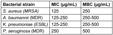
Figure 3: Comparative antimicrobial efficacy. Minimum Inhibitory Concentrations (MIC) of quantum dots against a panel of MDR pathogens.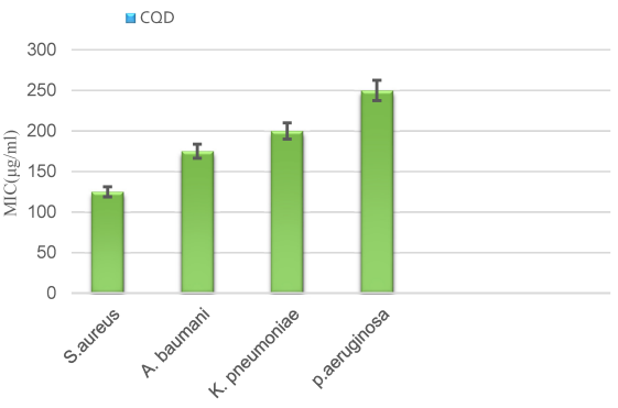
Down-regulation of biofilm-associated genes
To elucidate the mechanism of the anti-biofilm activity at the molecular level, real-time PCR (RT-qPCR) was performed to quantify the expression of key biofilm-related genes following exposure to sub-MIC concentrations (Figure 4 [Fig. 4]).
Figure 4: Comparative anti-biofilm efficacy. Dose-dependent inhibition of mature biofilms with CQD showing superior eradication capacity (>3.5 log10 CFU/mL reduction) at 4 times MIC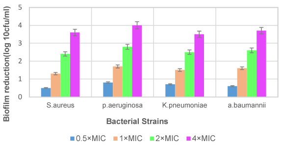
For S. aureus, expression of icaA and icaD (responsible for polysaccharide intercellular adhesin synthesis) was reduced by 4.5-fold and 5.2-fold, respectively (P<0.001). In P. aeruginosa, the lectin gene lecA was down-regulated 3.8-fold and the exopolysaccharide gene pelA by 4.1-fold (P<0.01). Similarly, K. pneumoniae showed reduced expression of fimbrial genes mrkA (3.5-fold) and mrkD (4.0-fold), while A. baumannii exhibited down-regulation of bap (biofilm-associated protein) and ompA (outer membrane protein A) by 4.2-fold and 3.9-fold, respectively. These results indicate that CQDs effectively disrupt biofilm formation at the transcriptional level.
In vivo wound healing efficacy
Over the 14-day observation period, topical treatment with CQD gel (1%) significantly accelerated the wound healing process compared to control groups (Figure 5 [Fig. 5], Figure 6 [Fig. 6]).
Figure 5: In vivo therapeutic evaluation in a murine polymicrobial infected wound model. Representative macroscopic images of infected wounds treated with different formulations over 14 days. The C QDs hydrogel group exhibited the most pronounced therapeutic effect, showing markedly accelerated wound contraction and tissue regeneration compared with the control, antibiotic-treated group, and metal-based quantum dots, including Ag QDs, Cu QDs, and ZnO QDs. Notably, by Day 8, wounds treated with C QDs showed substantial closure and reduced inflammation, whereas other treatment groups still displayed visible wound areas and delayed healing. By Day 14, the C QDs-treated wounds were nearly completely closed, indicating superior epithelial regeneration and overall healing efficacy.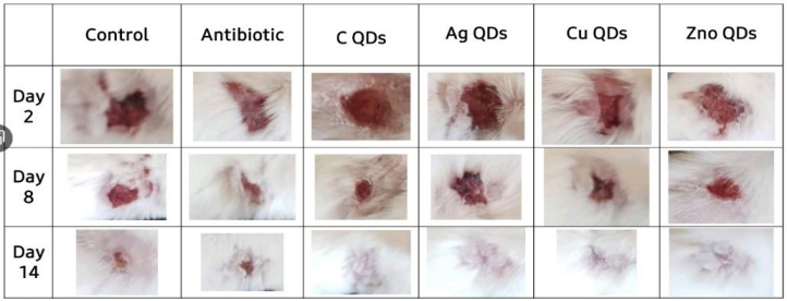
Figure 6: Quantitative analysis of in vivo wound closure rates over a 21-day period. The graph illustrates the percentage of wound contraction across three experimental groups: Uninfected Control, Standard Antibiotic, and Hydrogel Base. Data are presented as mean pm standard deviation (SD) [or SEM]. Statistical analysis indicates no significant difference between the groups at the specified endpoint (p = 0.48).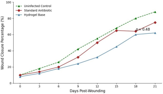
At day 7, wound contraction rates were 45.2±5.1% in the CQD-treated group, compared to 22.3±4.2% in the untreated control (P<0.01) and 38.5±4.8% in the antibiotic-treated group (P<0.05). By day 14, CQD-treated wounds achieved nearly complete closure (94.3±3.2%), whereas untreated controls remained at 68.5±5.6% (P<0.001). Histopathological analysis of tissue biopsies on day 14 revealed enhanced epithelialization, increased collagen deposition, and reduced inflammatory cell infiltration in CQD-treated wounds compared to controls (Figure 7 [Fig. 7]). Furthermore, bacterial load in wound tissue was significantly lower in the CQD group (log10 3.2±0.4 CFU/g) compared to untreated controls (log10 7.8±0.6 CFU/g) (P<0.001). Macroscopic images of wound healing progression and quantitative closure rates are presented in Figure 5 [Fig. 5], Figure 6 [Fig. 6], and Figure 7 [Fig. 7].
Figure 7: Efficacy of the experimental treatment on accelerated wound healing. Progression of wound closure (%) is compared between the Experimental Treatment and Control groups over a 21-day timeframe. The experimental group achieved a maximum closure rate of 95.3% by day 21. Delta symbols (Delta) denote a statistically significant difference between the experimental and control groups at corresponding time points (p < 0.05). Data are expressed as mean pm SD .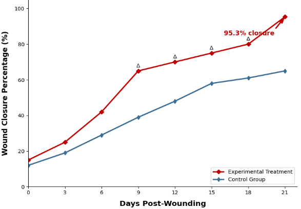
Discussion
The escalating global threat of AMR, particularly the growing challenge of biofilm-associated infections, necessitates the urgent development of novel therapeutic strategies [1], [3]. In this study, we successfully synthesized carbon quantum dots (CQDs) using a green, plant-based hydrothermal approach from oak fruit extract. The resultant CQDs demonstrated robust antimicrobial and anti-biofilm activities against major ESKAPE pathogens, culminating in significantly enhanced in vivo wound healing. Comparing the MIC with polihexanide, which is considered the first-line wound antiseptic for chronic wounds and burn wounds [32], the latter is far more effective than oak fruit CQDs (MIC for S. aureus and VISA strains 1 µg/mL, for E. coli 2 µg/mL) [33]. As consequence, oak fruit CQDs are likely to be less suitable for direct wound treatment and more suitable as an active ingredient in wound dressings.
A critical factor contributing to the exceptional efficacy of the synthesized CQDs is their unique physicochemical properties [12]. The green synthesis strategy not only provides an eco-friendly and cost-effective alternative to conventional chemical methods but also yields nanoparticles with optimal morphological traits [13], [14]. Our TEM analysis confirmed a spherical morphology with an ultra-small size of <10 nm (mean 6.8 nm). This sub-10 nm dimension is of paramount importance, as it enables the quantum dots to deeply penetrate the dense extracellular polymeric substance (EPS) matrix of bacterial biofilms, an obstacle that typically renders conventional antibiotics ineffective [6], [9]. Similar findings have been reported for CQDs derived from other biomass sources, such as orange peel and garlic, where particle sizes below 10 nm correlated with enhanced biofilm penetration [16], [17].
The in vitro results highlighted a dose-dependent bactericidal effect, with S. aureus emerging as the most susceptible strain (MIC=125 µg/mL). This high sensitivity makes these CQDs particularly promising for clinical applications, such as the development of advanced wound dressings for burn patients and intensive care unit (ICU) settings where S. aureus infections are highly prevalent [4], [5]. Conversely, P. aeruginosa exhibited the lowest inhibition (MIC=250–500 µg/mL), which aligns with its intrinsically robust outer membrane and highly adaptive efflux pump systems [7], [8]. This observation is consistent with previous reports on CQDs derived from other green precursors, where Gram-negative bacteria generally show lower susceptibility due to their additional outer membrane barrier [15].
Furthermore, our molecular analysis established a clear causal chain linking transcriptional regulation to phenotypic outcomes. The significant down-regulation of critical biofilm-associated genes—such as icaA/icaD in S. aureus and lecA/pelA in P. aeruginosa—under sub-MIC conditions indicates that CQDs disrupt biofilm formation at the genetic level [27], [28]. The ica operon is responsible for the synthesis of polysaccharide intercellular adhesin (PIA), a key component of staphylococcal biofilms [24]. Down-regulation of icaA and icaD by more than 4-fold suggests that CQDs interfere with PIA production, thereby reducing cell-to-cell adhesion. Similarly, pelA encodes an exopolysaccharide essential for the structural integrity of P. aeruginosa biofilms, and its suppression by 4.1-fold likely compromises biofilm architecture [26]. In K. pneumoniae, the down-regulation of mrkA and mrkD (type 3 fimbrial genes) is particularly noteworthy, as these genes mediate adherence to abiotic surfaces and host cells [5]. In A. baumannii, the reduced expression of bap (biofilm-associated protein) and ompA (outer membrane protein A) further supports the broad-spectrum anti-biofilm activity of CQDs [25].
This gene suppression translates directly to a diminished biofilm phenotype, which subsequently reduces the bacterial load within the infection site. Consequently, this molecular and phenotypic disruption provides a highly favorable microenvironment for tissue regeneration, elucidating the accelerated epithelialization and superior wound contraction observed in our in vivo murine model [29], [30]. Our in vivo results (94.3% wound closure at day 14) are comparable to or better than those reported for other nanoparticle-based wound dressings, including silver nanoparticles (typically 80–90% closure at day 14) [10], [11]. Importantly, CQDs are generally considered less cytotoxic than metallic nanoparticles (e.g., silver, copper, zinc oxide), which have been associated with dose-dependent toxicity in mammalian cells [12], [15]. This biocompatibility advantage makes CQDs particularly attractive for repeated topical application on open wounds.
While the exact molecular targets are still under investigation, our findings strongly suggest that CQDs interfere with biofilm formation at the transcriptional level. Several hypotheses exist, including
- generation of reactive oxygen species (ROS) leading to oxidative stress and membrane damage,
- physical interaction with bacterial cell walls causing membrane disruption, and
- internalization and interaction with nucleic acids or proteins [17], [20].
Our FTIR results confirmed the presence of surface carboxylic and hydroxyl groups, which are known to facilitate hydrogen bonding with bacterial surface proteins and polysaccharides, potentially disrupting cell signaling pathways involved in biofilm regulation [21]. Future studies should focus on identifying the precise molecular targets of CQDs using proteomic and metabolomic approaches.
Despite these highly promising findings, the translation of these nanoparticle-based therapeutics from bench to bedside requires further investigation. Future studies must prioritize comprehensive cytotoxicity evaluations using human cell lines (e.g., keratinocytes, fibroblasts) to ensure biocompatibility [13]. Additionally, detailed pharmacokinetic profiling (absorption, distribution, metabolism, excretion) and rigorous, controlled clinical trials are imperative to ascertain their safety and efficacy in human subjects [14]. From a pharmaceutical perspective, incorporating these green-synthesized CQDs into biocompatible hydrogel matrices (e.g., chitosan, alginate) could represent a highly effective strategy for developing sustained-release, smart wound dressings with both antimicrobial and wound-healing properties [30].
One limitation of the present study is the lack of long-term toxicity assessment beyond 14 days. Additionally, the potential for development of bacterial resistance upon repeated exposure to CQDs was not investigated. While some studies suggest that nanoparticles may have a lower propensity to induce resistance compared to conventional antibiotics, unlike conventional antiseptics where resistance development is not considered relevant [31], this remains an area requiring empirical validation for CQDs. Future studies should also explore the synergistic effects of CQDs in combination with conventional antiseptics, as such combinations could potentially lower the effective doses of both agents, reduce toxicity, and overcome existing resistance mechanisms.
Limitations
Infected wounds are generally treated topically with antiseptics rather than antibiotics. Chemotherapy is indicated only in cases where the infection has metastasized, in conjunction with topical wound antiseptic treatment [34]. In this context, the comparison group treated topically with ciprofloxacine does not adhere to the standard of antiseptic wound treatment, meaning that the results cannot be applied to the context of antiseptic wound treatment. Regardless, however, the oak fruit CQDs is antimicrobial effective. properties.
A major limitation is that it is not possible to assess whether the oak fruit CQDs meets the requirements for a wound antiseptic based solely on the MIC and MBC values. In Europe in the quantitative suspension test a wound antiseptic must achieve a ≥5 lg reduction of CFUs without organic challenges and ≥3 lg with organic challenges for acute wound antiseptics of 10% albumin or 10% sheep blood within a practical exposure time against S. aureus, P. aeruginosa, E. coli, E. faecium, and Candida albicans [35], or for conditions on chronic wounds as challenge Eagle’s minimal essential medium with Earle’s salts and L-glutamine, supplemented with 10% fetal calf serum without addition of antibiotics [36]. The next step of testing is the so-called phase 2/ step 2 test, a standardized in-vitro test on metal carrier with and without organic soil. The requirement to effectiveness is the same as in the quantitative suspension test [37], [38], [39].
Only once these requirements have been met will the next stages of testing follow, such as efficacy testing on experimentally contaminated 3D wound models and final the clinical trial [40].
Conclusion
The study demonstrates that hydrothermally synthesized oak-derived carbon quantum dots (CQDs) possess potent antimicrobial and anti-biofilm activity against a wide range of clinically relevant ESKAPE pathogens. The underlying mechanism involves the down-regulation of key biofilm-associated genes at the transcriptional level, leading to reduced biofilm formation and enhanced bacterial susceptibility. Furthermore, topical application of CQD-based gel significantly accelerated wound healing in an infected murine model, achieving near-complete wound closure within 14 days. These findings highlight the robust potential of green-synthesized CQDs as alternative therapeutic agents for managing multidrug-resistant wound infections. Future pharmacokinetic, toxicological, and clinical investigations are warranted to translate these promising preclinical results into advanced wound dressings for clinical use.
Notes
Authors’ ORCIDs
- Nabipour YS: https://orcid.org/0000-0003-0504-9586
- Hesampour A: https://orcid.org/0000-0002-7324-5469
- Asbchin SA: https://orcid.org/0000-0001-6079-1842
- Tajabadi M: https://orcid.org/0000-0003-2780-3447
- Rostamza A: https://orcid.org/0000-0002-2288-3794
Ethical approval
Animal experiments were conducted following the ethical guidelines approved by the Animal Ethics Committee of Tehran University of Medical Sciences (Ethics code: IR.TUMS.AEC.1402.089). All in vivo procedures were performed in accordance with the National Institutes of Health (NIH) Guide for the Care and Use of Laboratory Animals and the national guidelines of Iran for the ethical use of animals in research.
Funding
None.
This research received no specific grant from any funding agency in the public, commercial, or not-for-profit sectors.
Competing interests
The authors declare that they have no competing interests.
References
[1] Ferdinand AS, McEwan C, Lin C, Betham K, Kandan K, Tamolsaian G, Pugeva B, McKenzie J, Browning G, Gilkerson J, Coppo M, James R, Peel T, Levy S, Townell N, Jenney A, Stewardson A, Cameron D, Macintyre A, Buising K, Howden BP. Development of a cross-sectoral antimicrobial resistance capability assessment framework. BMJ Glob Health. 2024 Jan 16;9(1):e013280. DOI: 10.1136/bmjgh-2023-013280[2] Ruckert A, Lake S, Van Katwyk SR. Developing a protocol on antimicrobial resistance through WHO's pandemic treaty will protect lives in future pandemics. Global Health. 2024 Jan;20(1):10. DOI: 10.1186/s12992-024-01015-1
[3] Gaikwad S, Birla S, Ingle AP, Gade A, Ingle P, Golińska P, Rai M. Superior Wound-Healing Activity of Mycosynthesized Silver Nanogel on Different Wound Models in Rat. Front Microbiol. 2022;13:881404. DOI: 10.3389/fmicb.2022.881404
[4] Wilson MG, Pandey S. Pseudomonas aeruginosa Infections. Treasure Island (FL): StatPearls Publ; 2026 Jan. Available from: https://www.ncbi.nlm.nih.gov/books/NBK557831/
[5] Taylor TA, Tobin EH, Unakal CG. Staphylococcus aureus Infection. Treasure Island (FL): StatPearls Publ; 2026 Jan. Available from: https://www.ncbi.nlm.nih.gov/books/NBK441868/
[6] Grari O, Ezrari S, El Yandouzi I, Benaissa E, Ben Lahlou Y, Lahmer M, Saddari A, Elouennass M, Maleb A. A comprehensive review on biofilm-associated infections: Mechanisms, diagnostic challenges, and innovative therapeutic strategies. Microbe. 2025 Sept;8:100436. DOI: 10.1016/j.microb.2025.100436
[7] Høiby N, Bjarnsholt T, Givskov M, Molin S, Ciofu O. Antibiotic resistance of bacterial biofilms. Int J Antimicrob Agents. 2010 Apr;35(4):322-32. DOI: 10.1016/j.ijantimicag.2009.12.011
[8] Fazly Bazzaz BS, Khameneh B, Zarei H, Golmohammadzadeh S. Antibacterial efficacy of rifampin loaded solid lipid nanoparticles against Staphylococcus epidermidis biofilm. Microb Pathog. 2016 Apr;93:137-44. DOI: 10.1016/j.micpath.2015.11.031
[9] Ioannou P, Baliou S, Samonis G. Nanotechnology in the Diagnosis and Treatment of Antibiotic-Resistant Infections. Antibiotics (Basel). 2024 Jan 25;13(2):121. DOI: 10.3390/antibiotics13020121
[10] Nabipour YE, Hesampour A, Ahmady Asbchin S, Tajabadi M, Rostamzad A. Comparative study of the effects of biologically synthesized copper and zinc oxide quantum nanodots on biofilm-forming genes in Klebsiella pneumoniae and Staphylococcus aureus bacteria. Med Sci. 2026;36(2):189-99. DOI: 10.66224/iau.36.2.189
[11] Afrasiabi S, Partoazar A. Targeting bacterial biofilm-related genes with nanoparticle-based strategies. Front Microbiol. 2024;15:1387114. DOI: 10.3389/fmicb.2024.1387114
[12] Thejo Kalyani N, Dhoble SJ, Michalska-Domańska M, Vengadaesvaran B, Nagabhushana H, Arof AK. Quantum Dots. In: Woodhead Publishing Series in Electronic and Optical Materials. Woodhead Publishing;2023:3-40. DOI: 10.1016/B978-0-323-85278-4.00010-6
[13] Praseetha PK, Litany RIJ, Alharbi HM, Khojah AA, Akash S, Bourhia M, Mengistie AA, Shazly GA. Green synthesis of highly fluorescent carbon quantum dots from almond resin for advanced theranostics in biomedical applications. Sci Rep. 2024 Oct;14(1):24435. DOI: 10.1038/s41598-024-75333-0
[14] Gebre SH. Bio-inspired synthesis of metal and metal oxide nanoparticles: the key role of phytochemicals. J Clust Sci. 2023;34(2). DOI: 10.1007/s10876-022-02276-9
[15] Lou Y, Hao X, Liao L, Zhang K, Chen S, Li Z, Ou J, Qin A, Li Z. Recent advances of biomass carbon dots. Nano Sel. 2021;2:1117-45. DOI: 10.1002/nano.202000232
[16] Gul U, Kanwal S, Tabassum S, Gilani MA, Rahim A. Microwave-assisted synthesis of carbon dots as reductant and stabilizer for silver nanoparticles with enhanced-peroxidase like activity for colorimetric determination of hydrogen peroxide and glucose. Mikrochim Acta. 2020 Jan;187(2):135. DOI: 10.1007/s00604-019-4098-x
[17] Zhou Y, Liu Y, Li Y, He Z, Xu Q, Chen Y, Street J, Guo H, Nelles M. Multicolor carbon nanodots from food waste and their heavy metal ion detection application. RSC Adv. 2018 Jun;8(42):23657-62. DOI: 10.1039/c8ra03272f
[18] Bourang S, Jahanbakhsh Godehkahriz S, Asghari Zakaria R, Parsa H, Noruzpuor M. Green synthesis of iron oxide, copper, zinc oxide and silver nanoparticles from aqueous extract of F. vulgare and evaluation of their structural and antimicrobial properties. Agric Biotechnol J. 2024;16(3):60-88. DOI: 10.22103/jab.2024.23102.1557
[19] Liu T, Lu Y, Zhan R, Qian W, Luo G. Nanomaterials and nanomaterials-based drug delivery to promote cutaneous wound healing. Adv Drug Deliv Rev. 2023 Feb;193:114670. DOI: 10.1016/j.addr.2022.114670
[20] Shahbazi A, Dall E. Synthesis of quantum carbon dots (CQDs) from hard walnut skin by hydrothermal method. Amirkabir J Civ Eng. 2023;55(3):493-504. DOI: 10.22060/ceej.2023.21041.7614
[21] Sachdev A, Gopinath P. Green synthesis of multifunctional carbon dots from coriander leaves and their potential application as antioxidants, sensors and bioimaging agents. Analyst. 2015 Jun;140(12):4260-9. DOI: 10.1039/c5an00454c
[22] Sajadi M, Honarman A, Rahimi-Varposhti M, Yadegari L, Attarzadeh A, Mahjobipoor L, Fattahpour S. A comparison of disk diffusion method and E-test in determining the susceptibility and resistance of Klebsiella and Acinetobacter strains to cefepime in patients with ventilator-associated pneumonia admitted to the intensive care unit. Immunopathol Persa. 2022;8(2):e30330. DOI: 10.34172/ipp.2022.30330
[23] Rai S, Dash D, Agarwal N. Introducing the new face of CLSI M100 in 2023: An explanatory review. Indian J Med Microbiol. 2023;46:100432. DOI: 10.1016/j.ijmmb.2023.100432
[24] Ademakinwa OR, Adeyemo A, Ayodele SO, Nwamuo AS, Oluduro AO. Implications of biofilm-producing organisms among bacteria isolated from ear-, nose-, and throat-infected patients. Avicenna J Clin Microbiol Infect. 2024;11(3):100-6. DOI: 10.34172/ajcmi.3557
[25] Parvekar P, Palaskar J, Metgud S, Maria R, Dutta S. The minimum inhibitory concentration (MIC) and minimum bactericidal concentration (MBC) of silver nanoparticles against. Biomater Investig Dent. 2020 Jul;7(1):105-9. DOI: 10.1080/26415275.2020.1796674
[26] Davaeifar S, Shahabani-Zahiri H, Modarressi MH, Mohammadi M, Akbari-Noghabi K. Evaluation of the effect of zinc oxide nanoparticles on the inhibition of biofilm formation of standard pathogenic bacteria and comparison with drug resistant isolates. J Ilam Univ Med Sci. 2019;27(3):138-49. Available from: http://sjimu.medilam.ac.ir/article-1-5405-en.html
[27] Aflakian F, Hashemitabar G. Biosynthesized silver nanoparticles at subinhibitory concentrations as inhibitors of quorum sensing, pathogenicity, and biofilm formation in PAO1. Heliyon. 2025 Feb;11(4):e42899. DOI: 10.1016/j.heliyon.2025.e42899
[28] Costa C, Ferreira GD, Simões M, Silva JL, Campos MJ. Real-Time PCR Protocol for Detection and Quantification of Three Pathogenic Members of the Vibrionaceae Family. Microorganisms. 2022 Oct 18;10(10):2060. DOI: 10.3390/microorganisms10102060
[29] Zangeneh MM, Zangeneh A, Etemadi N, Tabatabaei Aghda SJ, Pourkamalzadeh M, Zand S, Abbaszadeh M, Asadzadeh R. The effect of nano silver particles of the aqueous extract of Artemisia aucheri on the healing process of skin wounds in rats. PBP. 2023;5(1):1-7. Available from: http://pbp.medilam.ac.ir/article-1-189-en.html
[30] Gaikwad S, Birla S, Ingle AP, Gade A, Ingle P, Golińska P, Rai M. Superior Wound-Healing Activity of Mycosynthesized Silver Nanogel on Different Wound Models in Rat. Front Microbiol. 2022;13:881404. DOI: 10.3389/fmicb.2022.881404
[31] Lauwereyns J, Bajramovic J, Bert B, Camenzind S, De Kock J, Elezović A, Erden S, Gonzalez-Uarquin F, Ulman YI, Hoffmann OI, Kitsara M, Kostomitsopoulos N, Neuhaus W, Petit-Demouliere B, Pollo S, Riso B, Schober S, Sotiropoulos A, Thomas A, Vitale A, Wilflingseder D, Ahluwalia A. Toward a common interpretation of the 3Rs principles in animal research. Lab Anim (NY). 2024 Dec;53(12):347-50. DOI: 10.1038/s41684-024-01476-2
[32] Kramer A, Dissemond J, Kim S, Willy C, Mayer D, Papke R, Tuchmann F, Assadian O. Consensus on Wound Antisepsis: Update 2018. Skin Pharmacol Physiol. 2018;31(1):28-58. DOI: 10.1159/000481545
[33] Assadian O, Wehse K, Hübner NO, Koburger T, Bagel S, Jethon F, Kramer A. Minimum inhibitory (MIC) and minimum microbicidal concentration (MMC) of polihexanide and triclosan against antibiotic sensitive and resistant Staphylococcus aureus and Escherichia coli strains. GMS Krankenhhyg Interdiszip. 2011;6(1):Doc06. DOI: 10.3205/dgkh000163
[34] Nabipour YS, Hesampour A, Ahmady Asbchin S, Tajabadi M, Rostamzad A. Differential Modulation of Biofilm-associated Gene Expression by Ag and ZnO Quantum Dots in Gram-positive and Gram-negative Bacteria: Implications for Cutaneous Wound Healing. Journal of Genetic Resources. 2025;11(2):268-76. DOI: 10.22080/jgr.2026.31027.1456
[35] Pitten FA, Werner HP, Kramer A. A standardized test to assess the impact of different organic challenges on the antimicrobial activity of antiseptics. J Hosp Infect. 2003 Oct;55(2):108-15. DOI: 10.1016/s0195-6701(03)00260-3
[36] Campbell KE, Keast D, Woodbury G, Houghton P. Wear time in two hydrocolloid dressings using a novel in-vivo-model. Wounds. 2003;15:40-8.
[37] Igelbrink D, Koburger T, Simon A, Koburger T, Kramer A. Microbiocidal efficacy of MedihoneyTM. GMS Krankenhaushyg Interdiszip. 2007;2(2):Doc50.
[38] Ebert M, Assadian O, Hübner NO, Koburger T, Kramer A; International Society of Chemotherapy Working Group Antiseptics; Working Section for Clinical Antiseptic of the German Society for Hospital Hygiene. Antimicrobial efficacy of the silver wound dressing Biatain Ag in a disc carrier test simulating wound secretion. Skin Pharmacol Physiol. 2011;24(6):337-41. DOI: 10.1159/000330761
[39] Schedler K, Assadian O, Brautferger U, Müller G, Koburger T, Classen S, Kramer A. Proposed phase 2/ step 2 in-vitro test on basis of EN 14561 for standardised testing of the wound antiseptics PVP-iodine, chlorhexidine digluconate, polihexanide and octenidine dihydrochloride. BMC Infect Dis. 2017 Feb;17(1):143. DOI: 10.1186/s12879-017-2220-4
[40] Besser M, Dietrich M, Weber L, Rembe JD, Stuermer EK. Efficacy of antiseptics in a novel 3-dimensional human plasma biofilm model (hpBIOM). Sci Rep. 2020 Mar;10(1):4792. DOI: 10.1038/s41598-020-61728-2