
Digital versus conventional impression-taking of the ear canal: A comparison of accuracy between a handheld scanner and impression material
Henrik Holger Mallwitz 1Vitalij Joukov 2
Joachim Albrecht 2
Steffen Kreikemeier 1
1 Center of Excellence in Audiology, Aalen University of Applied Sciences, Aalen, Germany
2 Research Institute for Innovative Surfaces (FINO), Hochschule Aalen, Germany
Abstract
Background: The comfort of hearing aids is directly linked to the fitting of the earmolds. Additive manufacturing is now standard practice in the production of earmolds, which is why digitally capturing the ear canal is the critical first step. Traditionally, this is done using silicone impression materials, followed by scanning the impressions. A handheld scanner or ear canal scanner for direct digital capture has recently become available on the market as an alternative. The aim of this study is to evaluate the accuracy of this digital system in comparison with the conventional method.
Method: Two ear canal models were produced using 3D-printing technology to serve as reference models. Five users each took three conventional and three digital impressions. The conventional impressions were digitised using a desktop scanner. The scans were compared with the reference model using software-assisted best-fit superimposition. The mean of the deviation and the standard deviation of the deviation were evaluated.
Results: Digital impression-taking resulted in a lower mean of the deviation (–0.052 mm) compared with impression materials (Material 1: –0.136 mm; Material 2: –0.118 mm), suggesting slight shrinkage of the impression materials. The standard deviations of the deviation were 0.415 mm for the handheld scanner and between 0.496 mm and 0.546 mm for the conventional methods. The influence of geometry or the user was minimal.
Conclusion: Digital impression-taking using a handheld scanner is an equivalent alternative to conventional impression-taking. The advantages lie primarily in the workflow, time savings and the patient experience, whilst the high initial costs are currently limiting its widespread adoption.
Keywords
handheld scanners, ear canal scanners, impression material, ear impressions, additive manufacturing, target-actual comparison, earmolds
Introduction
The comfort of a hearing aid is primarily ensured by a good fit of the earmold. Nowadays, most earmolds are manufactured using 3D-printing technology [1], [2]. Therefore, the first step in the production of an earmold is to obtain a digital image of the ear canal. Traditionally, this is achieved by taking an impression of the ear canal using a special silicone impression material, followed by scanning the impression. A device made by Natus has recently become available on the market that allows the geometry of the ear canal to be scanned and digitised directly in 3D. This handheld scanner, known as the ‘Otoscan’, offers advantages in terms of time savings, material costs and patient comfort. At the same time, this method is limited by high initial costs, whereas the conventional method using impression material, although more labour-intensive, is well-established and more cost-effective [3]. To date, there have been few studies that systematically compare the accuracy of the two methods. Zheng et al. and Mølhave et al. compare the subjective wearing comfort of earmolds produced using data from both the conventional and the direct digital method [3], [4]. The aim of this study is to compare the two methods objectively, using a reference object, in order to evaluate the current state of the art of the available handheld scanner and to provide a well-founded recommendation to the specialist audience.
Figure 1 [Fig. 1] shows, on the left, the scanning process using the ear canal scanner. The measuring probe, which incorporates a ring laser, an LED and a camera, is moved along the patient’s ear canal and the contours of their ear. The progress of the scanning process can be monitored live on the screen. It shows how the scanned data is being assembled into a digital image of the ear in real time. The coded ring around the patient’s ear serves as a reference for the device to determine its relative position in relation to the patient’s ear. The resulting digital image of the ear canal can be seen on the right in Figure 1 [Fig. 1].
Figure 1: Scanning process using the handheld scanner (left) and the resulting digital image of the ear canal (right)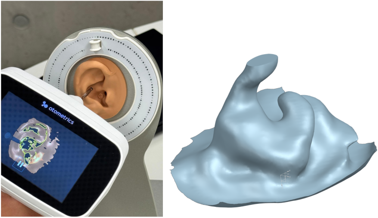
Methods
As a reference for analysing the differences between digital and conventional impressions, two artificial ear canals (one straight and angular, the other anatomically curved) were designed using a computer-aided design (CAD) system (Siemens NX 1980) and attached to an existing 3D-model of an ear. The two ears and ear canals were manufactured using a stereolithography 3D-printer (Asiga PRO2) with the Composer 2.0.4 slicer software. Stereolithography (SLA) is an additive manufacturing process that uses an ultraviolet (UV) laser to cure liquid photopolymer resin into plastic components. The objects are built up layer by layer by projecting the cross-section of the 3D-model onto the resin [5]. A skin-coloured resin (pro3dure printodent GR-13) was used to provide the optical system with a realistic surface. Figure 2 [Fig. 2] shows, on the left, the reconstructed ear with the anatomical ear canal. The ring-shaped surface around the artificial ear serves as a contact point for the coded ring of the handheld scanner. On the right in Figure 2 [Fig. 2] is the finished skin-coloured 3D-printed model of the ear.
Figure 2: Design of the artificial ear canal, with the 3D-printed model to the right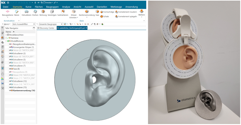
In addition to the handheld scanner (Natus Otoscan version 1.7.12883.0), impression materials from two different manufacturers (egger A/soft and pro3dure otosil IS-2) were compared. A total of five investigators took part in the test. Each person took three conventional impressions for each manufacturer, as well as three digital impressions using the handheld scanner. For the conventional method, a desktop scanner (smart optics mono Scan) was used to digitise the impressions. A total of 90 scans were recorded. Figure 3 [Fig. 3] shows, on the right, a scan of an impression of the anatomical ear canal taken using impression material made by pro3dure. Left in Figure 3 [Fig. 3] is the ‘ideal’ impression generated by the CAD system. To achieve this, the existing CAD volume model was converted into a surface model and cropped so that only the relevant area can be compared.
Figure 3: Ideal impression of the straight ear canal (left) and a scan of an impression taken with impression material (right)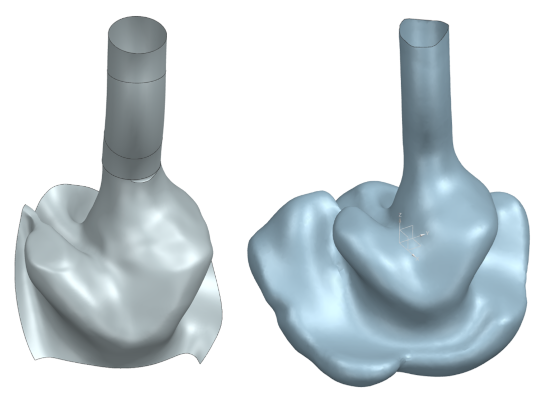
The resulting 3D models were compared with the surface model of the reference ear canal using computer-assisted best-fit registration (Geomagic Design X 2024 2.0). Before the comparison, the scan and the reference were aligned with one another (see Figure 4 [Fig. 4]), revealing initial differences between the scan (green) and the reference (blue). The Alignment is a crucial step prior to the actual comparison, as incorrect alignment would distort the measurement results. Challenges arose with individual datasets due to non-alignable models. In this case, the test subjects had to take additional impressions.
Figure 4: Scan and reference superimposed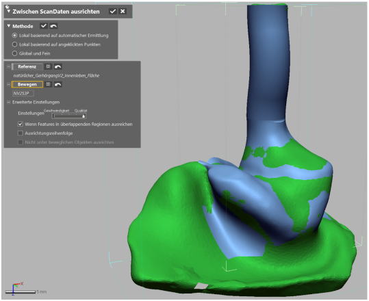
Once alignment was complete, the distances between the scan and the reference were compared and determined. Figure 5 [Fig. 5] shows the dimensional deviations in the scan of the impression highlighted in colour. The areas of the impression’s surface coloured yellow or red are higher than the reference, whilst the blue areas are lower. The mean of the deviation, which indicates shrinkage, was evaluated, as was the standard deviation of the deviation as a direction-independent measure of deviation.
Figure 5: Dimensional deviations in the scan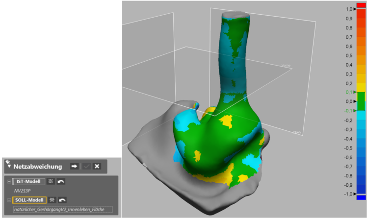
Results
Both methods demonstrate comparable overall accuracy. The difference in accuracy between the various impression materials is negligible. The mean of the deviation and the standard deviation of the deviation were calculated as measures of accuracy.
Table 1 [Tab. 1] shows the average values for all comparisons. Digital impressions exhibit a lower mean of the deviation. The standard deviation of the deviation for digital impressions is also slightly lower than that for impression materials. Differences between the five users and the two different artificial ear canals fall within a similarly narrow range (see Table 2 [Tab. 2] and Table 3 [Tab. 3], using the values for conventional impressions as an example).
Table 1: Results of the comparison between digital and conventional impression-taking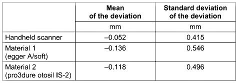
Table 2: Comparison of the two different types of artificial ear canals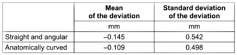
Table 3: Comparison of the accuracy achieved by different individuals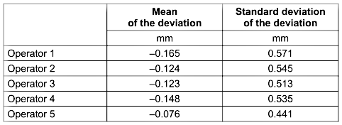
Table 2 [Tab. 2] compares the accuracy achieved with the two differently designed ear canals used. The values are slightly higher for the straight, angular ear canal. Table 3 [Tab. 3] shows the accuracy of the five different users. Operator 5, a particularly experienced user, represents an outlier.
Conclusions
It is important to note that the obtained values are not absolute, as the unknown manufacturing tolerance of the SLA 3D printer must theoretically be taken into account. However, the values are comparable, as the same printed artificial ear was used for all impressions. Digital impression-taking is already a viable alternative to conventional impression-taking. Both methods currently offer comparable precision. With both methods, slightly higher deviations are observed in the case of a straight, angular ear canal. This is likely due to the fact that the scanners used tend to smooth out sharp edges. The increased average deviation of the deviation observed with the conventional method suggests a slight shrinkage of the impression materials. This can be eliminated during digital post-processing. With the conventional method, air bubbles also contributed to increased deviations in some cases. With the digital method, however, incomplete scanning can lead to gaps in the data [6]. The key differences between the two methods therefore lie less in accuracy than in (acquisition) costs, workflow and user experience: the digital method is faster and offers patients an added value in terms of the experience, as the scanning process can be viewed in real time.
Future research
Plans include examinations of real ear canals and further tests involving additional users to capture inter-individual variability. For example, the results from beginners, advanced users and professional users will be compared. New handheld scanner models are expected in the third quarter of 2026, and preparations are already underway to test them. It is also conceivable that further impression materials and desktop scanners will be tested and compared. In addition, various factors affecting the accuracy of conventional impressions are to be investigated, such as cooling, heating or UV treatment of the impressions, as well as the presence of hair and earwax.
Notes
Conference presentation
This contribution was presented at the 28th Annual Conference of the German Society of Audiology and published as an abstract [7].
Funding
This work was supported by the KMU-innovativ program, funded by the German Federal Ministry of Research, Technology and Space (BMFTR), funding code 13XP5223B.
Competing interests
The authors declare that they have no competing interests.
References
[1] Sandström CG. The non-disruptive emergence of an ecosystem for 3D Printing: Insights from the hearing aid industry's transition 1989–2008. Technological Forecasting and Social Change. 2016 Jan;102:160-68. DOI: 10.1016/j.techfore.2015.09.006[2] Berufsgenossenschaft Holz und Metall. Schutzwirkung von Gehörschutz-Otoplastiken: Aktueller Technik- und Wissensstand sowie Studienergebnisse zu den Einflüssen von Fertigung und Handhabung. Mainz: Berufsgenossenschaft Holz und Metall; 2017 Nov.
[3] Zheng Y, Didion K, Ferguson N. Comparison of Traditional Impression and 3D Ear Scanning Techniques, Earmold Comfort, and Audiology Clinical Implications: A Pilot Study. Bioengineering. 2023 Dec 16;10(12):1431. DOI: 10.3390/bioengineering10121431
[4] Mølhave M, Madsen M, Degnbol LM, Christiansen ALB, Ovesen T. Three-dimensional scan versus conventional mould of outer ear in fabricating earmolds for hearing aids: a randomized controlled trial. International Journal of Audiology. 2026 Feb 18:1-8. DOI: 10.1080/14992027.2026.2630118
[5] Devi KS, Nilanjana R. Additive Manufacturing. Bengaluru: Trinity Press; 2022.
[6] Kreikemeier S, Mallwitz HH. Reproduzierbarkeit des Gehörgangscans bei unterschiedlich großer Detailaufnahme. In: Deutsche Gesellschaft für Audiologie e. V., editor. 28. Jahrestagung der Deutschen Gesellschaft für Audiologie. Oldenburg, 04.-06.03.2026. Düsseldorf: German Medical Science GMS Publishing House; 2026. Doc232. DOI: 10.3205/26dga232
[7] Mallwitz HH, Kreikemeier S, Albrecht J, Joukov V. Digitale versus konventionelle Abformung des Gehörgangs: Ein Genauigkeitsvergleich zwischen Handscanner und Abformmasse. In: Deutsche Gesellschaft für Audiologie e. V., editor. 28. Jahrestagung der Deutschen Gesellschaft für Audiologie. Oldenburg, 04.-06.03.2026. Düsseldorf: German Medical Science GMS Publishing House; 2026. Doc157. DOI: 10.3205/26dga157