
Single-stage multilamellar upper eyelid reconstruction after traumatic avulsion injury from a dog bite
Bernardo Ribeiro Cavadas 1Inês Oliveira Pires 1
Sara Alves 2,3
Ana Maria Magriço 4
Diogo Pais 3
Diogo Casal 1,3
1 Plastic Surgery Department and Burn Unit, Unidade Local de Saúde de São José, Lisbon, Portugal
2 Pathology Department, Unidade Local de Saúde de São José, Lisbon, Portugal
3 Anatomy Department, NOVA Medical School, Lisbon, Portugal
4 Ophthalmology Department, Unidade Local de Saúde de São José, Lisbon, Portugal
Abstract
Objective: To report a rare case of complete multilamellar avulsion of the upper eyelid following a dog bite, successfully managed with a single-stage reconstruction.
Methods: After meticulous debridement, single-stage reconstruction was performed using a lateral V-Y myocutaneous advancement flap, superior levator palpebrae superioris and Müller’s muscle flaps, and a composite tarsoconjunctival-skin graft prepared from the amputated tissue.
Results: At five years postoperatively, the patient demonstrated excellent functional and aesthetic outcomes, including full eyelid mobility, normal ocular surface protection, and symmetric eyelid contour without retraction, ectropion, or entropion.
Conclusions: Meticulous single-stage multilamellar reconstruction, including reuse of avulsed tissue after debridement, can restore critical eyelid function and aesthetics in contaminated trauma, avoiding staged procedures and donor site morbidity.
Keywords
eyelids, reconstructive surgical procedures, bites and stings, surgical flaps, traumatic amputation
Introduction
The eyelids are complex, dynamic structures essential for protecting the eye and preserving vision [1], [2]. The upper eyelid plays a crucial role in corneal protection and ocular surface lubrication [3]. Traumatic eyelid avulsion with full-thickness loss of all three lamellae is extremely rare and represents a significant reconstructive challenge [4], [5]. Dog bites are a common cause of facial trauma, but complete multilamellar eyelid avulsion is particularly infrequent and raises concerns regarding contamination and tissue viability [6]. Traditionally, large full-thickness eyelid defects require staged procedures such as the Cutler-Beard flap, which carry donor site morbidity and potential functional limitations [7], [8]. Ideally, amputated segments should be replanted using microvascular anastomoses; however, eyelid vessels are extremely small, and documented cases of successful replantation are extremely rare [9]. We present a rare case of complete multilamellar avulsion of the upper eyelid following a dog bite, successfully managed with a single-stage reconstruction using local flaps and a composite graft from the avulsed tissue.
Case description
A 20-year-old female with no prior medical conditions, allergies, or immunosuppression, and up-to-date vaccinations, presented to the emergency department following a dog bite to the right upper eyelid. The injury resulted in complete avulsion of the middle two-thirds of the upper eyelid involving all three lamellae. The amputated tissue was brought in and preserved under appropriate conditions in saline-soaked gauze. Ophthalmologic assessment excluded acute ocular injuries, and the globe remained intact (Figure 1 [Fig. 1] and Figure 2 [Fig. 2]).
Figure 1: Preoperative photograph showing complete avulsion of the middle two-thirds of the right upper eyelid following dog bite. The globe remains intact without acute injury. 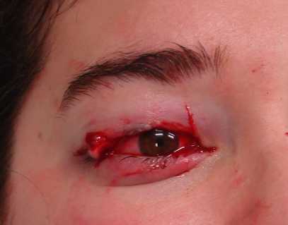
Figure 2: Intraoperative photograph of the amputated upper eyelid tissue immediately after receipt in the operating room. A ruler is included for scale reference. The tissue was preserved and used for composite graft reconstruction. 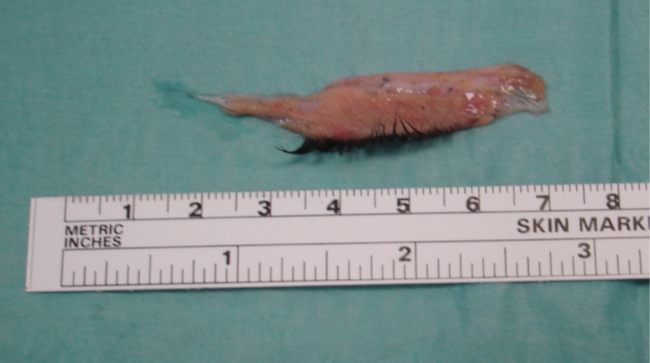
The patient was taken to the operating room within 6 hours of the trauma. Under general anesthesia, meticulous wound irrigation and debridement were performed to reduce the risk of infection. After thorough examination of the avulsed segment under the operating microscope, no vessels suitable for microvascular anastomoses were found. Devitalized edges of the amputated tissue were carefully trimmed. To facilitate reconstruction, the avulsed eyelid segment was incised in three layers along the margin, as shown in Figure 3 [Fig. 3]: the anterior layer (skin and subcutaneous tissue), the middle layer (orbicularis muscle), and the posterior layer (tarsal plate and palpebral conjunctiva).
Figure 3: Sagittal section of cadaveric upper eyelid stained with Masson’s trichrome, original magnification x10, demonstrating the trilayer anatomy. The anterior lamella includes skin (1), subcutaneous fat (2), and orbicularis oculi muscle (3). The middle lamella includes pre-septal fat (4), orbital septum (5), and post-septal fat pads (6). The posterior lamella comprises the tarsal plate (7) and palpebral conjunctiva (8). Upper eyelid specialized structures are visible: tarsal (meibomian) glands (9), sebaceous glands of eyelashes (Zeis glands) (10), apocrine glands (Moll glands) (11), and accessory lacrimal glands (Wolfring glands) (12). Eyelashes (black arrowheads), peripheral superior palpebral arterial arcade (yellow arrowhead), and marginal superior palpebral artery (asterisk) are noted. Red and blue dotted lines represent surgical incisions used to divide the amputated eyelid into layers for vascularization. 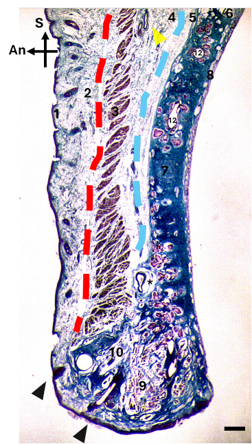
An, anterior; S, superior. Scale bar = 1 mm.
Initially, a superior lateral canthotomy was performed to allow mobilization of the lateral aspect of the remaining eyelid. A lateral V-Y myocutaneous advancement flap was mobilized medially, with the orbicularis muscle of this flap positioned between the subcutaneous tissue and orbicularis muscle of the amputated segment. The superior levator palpebrae superioris and Müller's muscle from the upper margin of the wound were mobilized inferiorly and sutured into the gap between the orbicularis muscle and the tarsal plate/conjunctiva of the avulsed eyelid.
The free margins of the bulbar and palpebral conjunctiva were sutured together with an inverted running 7/0 polyglactin 910 suture (Vicryl, Ethicon Inc., CAS: 26780-50-7). The flaps and composite graft were carefully inset to restore eyelid height, contour, and lamellar integrity, ensuring functional closure and ocular surface protection. The flaps were secured with interrupted 6/0 polyglactin 910 sutures (Figure 4 [Fig. 4]).
Figure 4: Immediate postoperative photograph demonstrating reconstruction using V-Y myocutaneous advancement flap and composite graft inset. Sutures are visible at the reconstruction site showing multilayer restoration.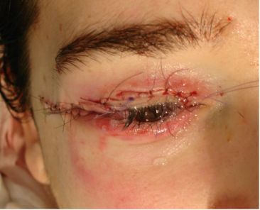
Postoperatively, the eyelid was dressed for 3 days with chloramphenicol eye ointment (CAS: 56-75-7) and petroleum jelly gauze. The wounds were exposed on postoperative day 4, irrigated with sterile saline, and treated with topical ointment three times daily for 15 days. The patient was discharged on postoperative day 8 with unremarkable recovery.
The composite graft showed good integration, and surgical wounds healed appropriately. At five years postoperatively, the patient maintained excellent eyelid mobility with full opening and closing, normal ocular surface protection without lagophthalmos, and no episodes of keratitis or ocular complications. The reconstructed eyelid preserved adequate contour and symmetry, with an inconspicuous scar (Figure 5 [Fig. 5]).
Figure 5: Long-term follow-up photograph at five years postoperatively demonstrating excellent functional and aesthetic outcome with symmetric eyelid contour, full mobility, and inconspicuous scar. No retraction, lagophthalmos, ectropion, or entropion present.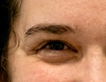
Discussion
Traumatic avulsion of the upper eyelid with full-thickness loss of all three lamellae represents one of the most challenging oculoplastic reconstructive problems [4], [5]. The presence of contamination from a dog bite adds significant concern regarding tissue viability and infection risk [6].
Traditionally, for large full-thickness upper eyelid defects, reconstructive options depend on defect size and extent. Direct primary closure is feasible only for small defects. Larger defects typically require local flaps, skin grafts, or combined techniques. For extensive defects involving all lamellae, staged procedures such as the Cutler-Beard flap have historically been the standard approach, though these require a second stage, carry donor site morbidity (particularly from the contralateral upper eyelid), and may result in functional limitations [7], [8]. Alternative approaches include free tissue transfer or use of distant grafts; however, these do not provide optimal “like-for-like” replacement of eyelid tissue [10].
The key advantage of the single-stage approach used in this case was the ability to restore all three lamellae in one operation while preserving the physiologic and structural properties of native eyelid tissue. By meticulously debriding the avulsed segment, removing devitalized tissue, and preparing it as a composite graft – subdividing it into its constituent layers and inlaying them with appropriate vascularization from local flaps – we achieved functional multilayer restoration. This avoided donor site morbidity from contralateral tarsus, auricular cartilage, or hard palate grafts.
A critical point in this case was that despite the contaminated nature of the dog bite injury, meticulous debridement and preparation of the amputated tissue allowed for its safe and effective reuse. The long-term follow-up demonstrates the durability of this strategy, with the patient maintaining full eyelid mobility, adequate corneal protection, and aesthetic symmetry without retraction, ectropion, or entropion at five years. In this patient, the favorable outcome indicates that the reuse of avulsed tissue from a contaminated injury can be a successful strategy when combined with meticulous debridement and vascularized flap coverage, potentially offering an alternative to staged procedures in similar clinical scenarios.
Limitations of this approach include the following:
- this is a single case report and does not provide comparative data to draw generalizable conclusions;
- the technique requires relatively well-preserved tissue suitable for composite grafting;
- success depends on early presentation and operative intervention;
- meticulous debridement and layering require expertise in oculoplastic surgery and microsurgical technique; and
- long-term follow-up data beyond this single case are needed to determine reproducibility and frequency of complications.
Although technically straightforward, this surgical option requires further validation through case series or comparative studies from other authors and surgical centers.
Conclusion
This case demonstrates that single-stage multilamellar reconstruction using local flaps and a composite graft from avulsed tissue can achieve excellent outcomes. However, the success of this approach likely depends on factors such as early presentation, availability of relatively well-preserved tissue, and the use of vascularized local flaps.
Notes
Ethics
The patient provided written informed consent for the publication of this case report and accompanying images. This report adhered to the ethical principles outlined in the Declaration of Helsinki as amended in 2013.
Competing interests
The authors declare that they have no competing interests.
References
[1] DiFrancesco LM, Codner MA, McCord CD. Upper eyelid reconstruction. Plast Reconstr Surg. 2004 Dec;114(7):98e-107e. DOI: 10.1097/01.prs.0000142743.57711.48[2] Soueid NE, Khoobehi K, Lee MR. Microsurgical replantation of total upper eyelid avulsion. Ann Plast Surg. 2006 Jan;56(1):99-102; discussion 102-3. DOI: 10.1097/01.sap.0000188106.44691.3d
[3] Espinoza GM, Prost AM. Upper Eyelid Reconstruction. Facial Plast Surg Clin North Am. 2016 May;24(2):173-82. DOI: 10.1016/j.fsc.2015.12.007
[4] Codner MA, McCord CD, Mejia JD, Lalonde D. Upper and lower eyelid reconstruction. Plast Reconstr Surg. 2010 Nov;126(5):231e-245e. DOI: 10.1097/PRS.0b013e3181eff70e
[5] Morley AM, deSousa JL, Selva D, Malhotra R. Techniques of upper eyelid reconstruction. Surv Ophthalmol. 2010 May-Jun;55(3):256-71. DOI: 10.1016/j.survophthal.2009.10.004
[6] Rafii AA, Enepekides DJ. Upper and lower eyelid reconstruction: the year in review. Curr Opin Otolaryngol Head Neck Surg. 2006 Aug;14(4):227-33. DOI: 10.1097/01.moo.0000233592.76552.d2
[7] Pham RT. Reconstruction of the upper eyelid. Otolaryngol Clin North Am. 2005 Oct;38(5):1023-32. DOI: 10.1016/j.otc.2005.03.014
[8] Stein JD, Antonyshyn OM. Aesthetic eyelid reconstruction. Clin Plast Surg. 2009 Jul;36(3):379-97. DOI: 10.1016/j.cps.2009.02.011.
[9] Yazici B, Ozturker C, Cetin Efe A. Reconstruction of Large Upper Eyelid Defects With Bilobed Flap and Tarsoconjunctival Graft. Ophthalmic Plast Reconstr Surg. 2020 Jul/Aug;36(4):372-374. DOI: 10.1097/IOP.0000000000001557
[10] Poh EW, O’Donnell BA, McNab AA, Sullivan TJ, Gaskin B, Malhotra R, Ataullah S, Cannon PS, Leatherbarrow B, Chan WO, Davis G, Selva D. Outcomes of upper eyelid reconstruction. Ophthalmology. 2014 Feb;121(2):612-3.e1. DOI: 10.1016/j.ophtha.2013.10.010